NEWS

05/03/2024
A groundbreaking study published by a USC-led international consortium offers new insights into stroke recovery, revealing that age-related changes in brain white matter significantly affect how well individuals recover motor abilities following a stroke.
04/23/2024
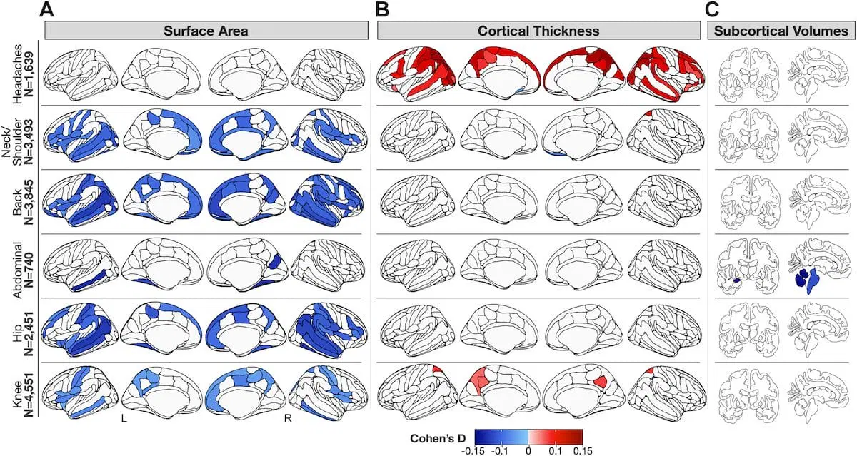
A new neuroimaging study pinpoints differences in gray matter in those with chronic pain, especially multisite pain, compared to healthy individuals. Those who reported head pain showed unique differences compared to other chronic pain types.
04/16/2024
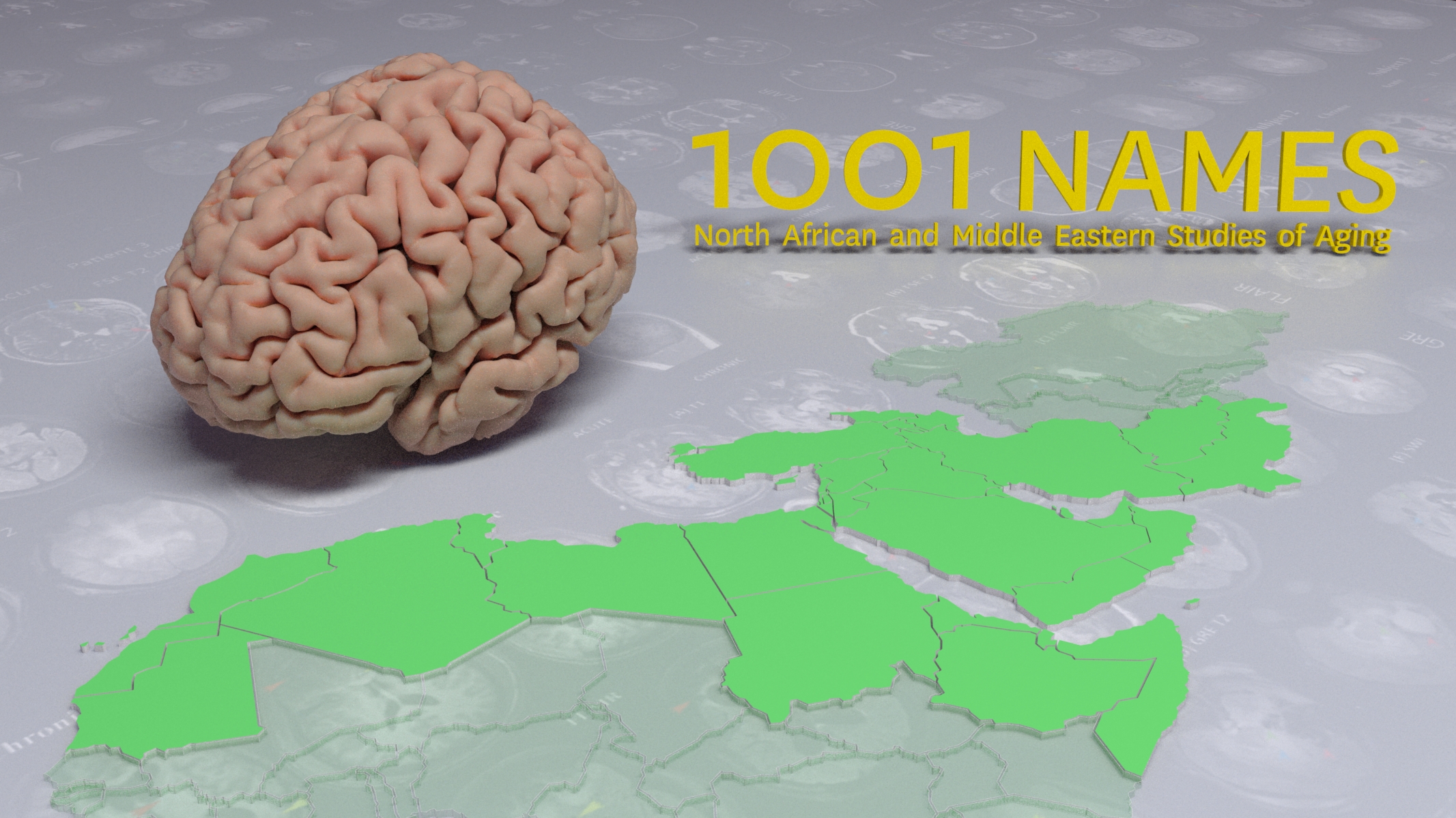
A new study led by Neda Jahanshad, PhD, is set to illuminate the underexplored domain of brain aging and risk for Alzheimer’s disease and related dementias (ADRD) among Middle Eastern and North African (MENA) adults in the US.
02/14/2024
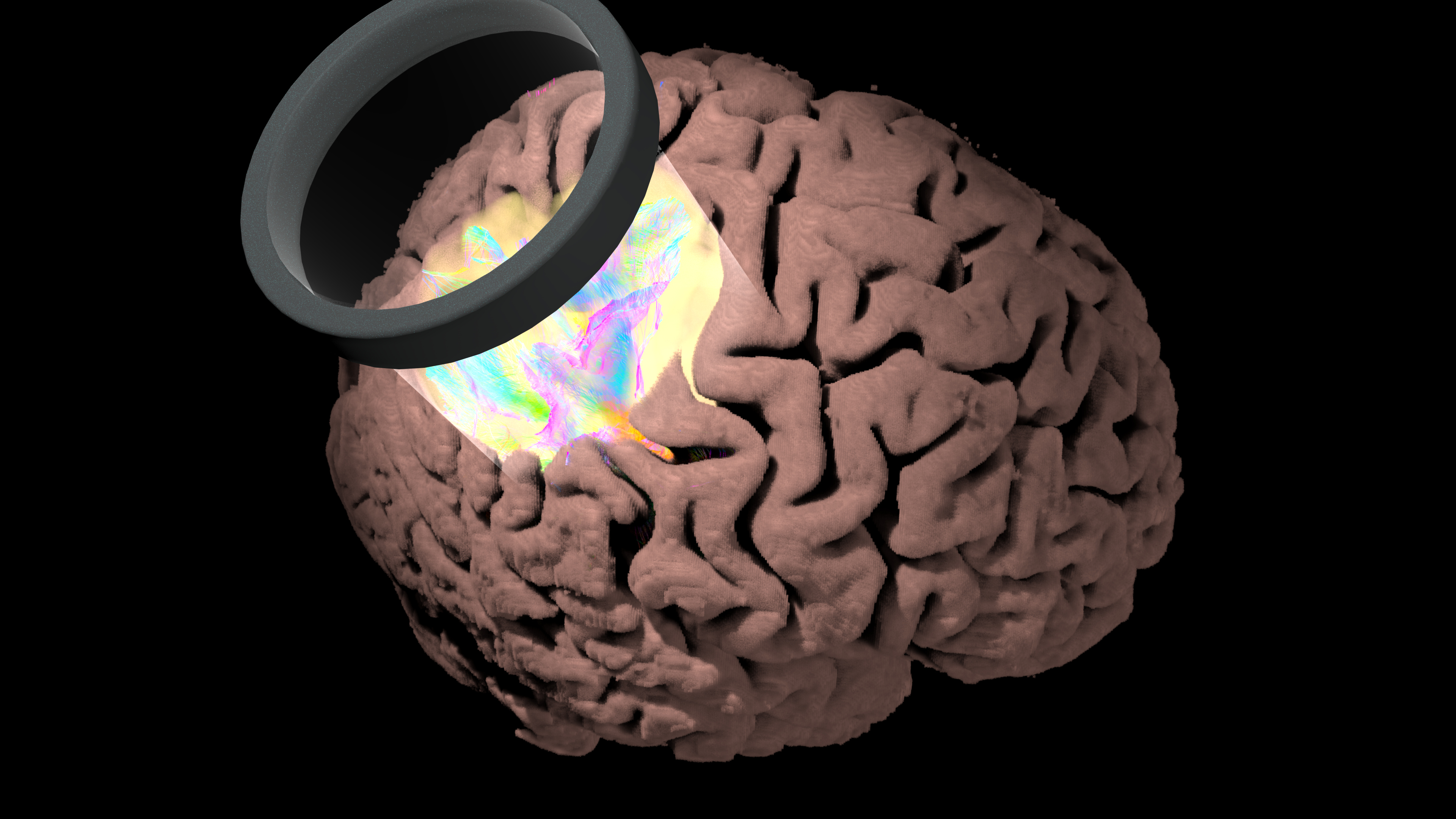
Uniting experts in artificial intelligence, psychiatry, and neuroimaging, the ENIGMA Consortium will apply AI to brain scans of people with depression in the first global analysis for new ways to predict which patients will respond best to a promising new treatment.
11/29/2023
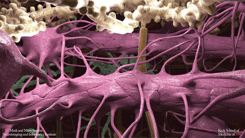
Recent breakthroughs suggest new ways to prevent, diagnose, and treat brain disorders associated with aging, including Alzheimer’s disease and related dementias
09/07/2023
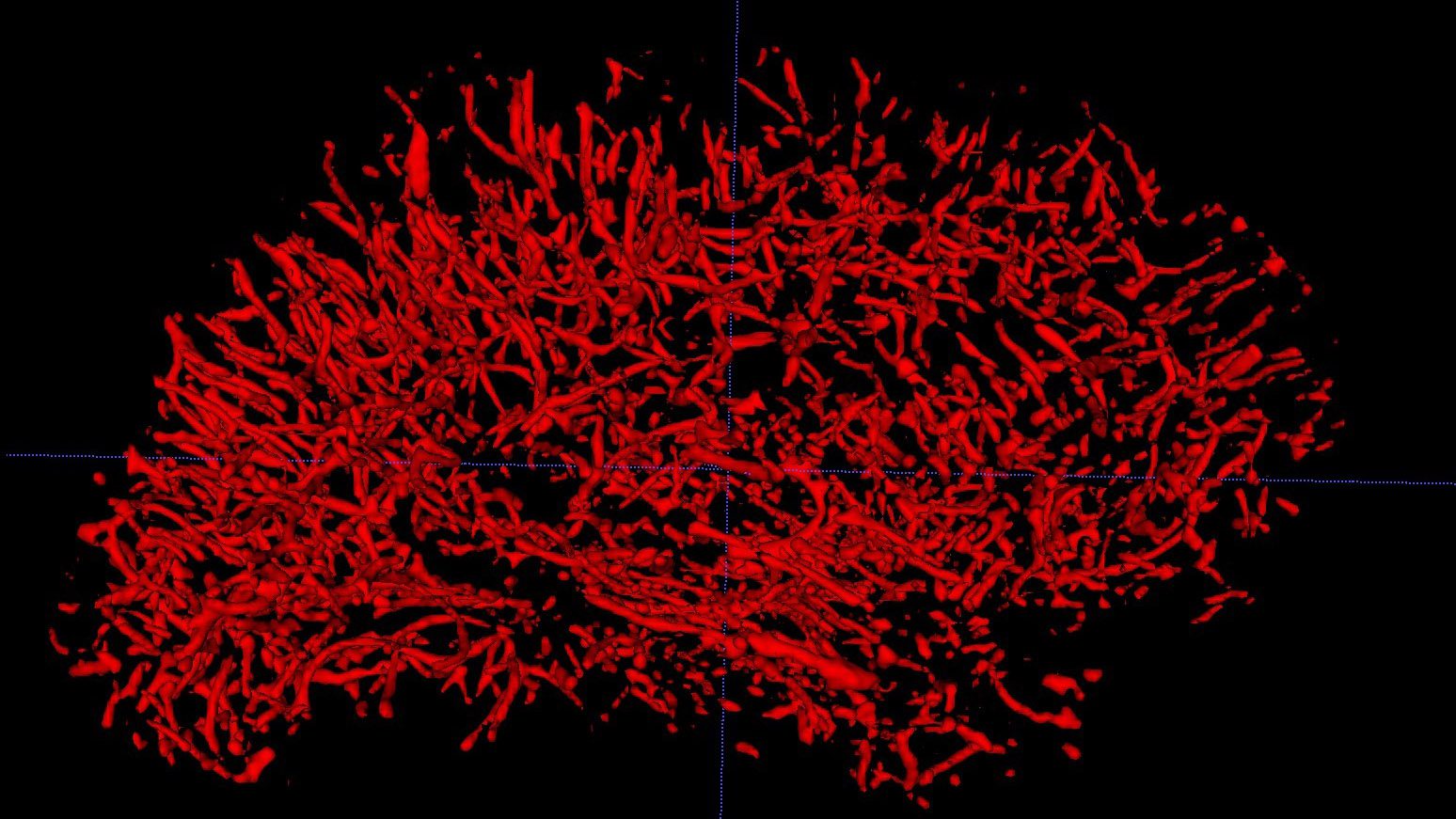
The Stevens INI expands their suite of health disparities research with a new award to examine vascular causes of dementia in Asian Americans using neuroimaging.

05/09/2023
Melody LaTorre was pregnant when she learned she had brain cancer. In a race against time, a team of Keck Medicine of USC experts worked to save both mother and child.

04/18/2023
USC Stevens INI releases their interactive 2022 annual report

04/06/2023
A USC-led team of researchers find that brain age, a neuroimaging-based assessment of global brain health, may play a role in post-stroke outcomes and could potentially help identify people at risk for poorer outcomes.

02/21/2023
As part of the ENIGMA Consortium, the India ENIGMA Initiative for Global Aging and Mental Health is a worldwide coordinated study of brain aging and Alzheimer’s disease to identify predictive markers in the blood, genome, and epigenome, and to better understand prognosis while supporting personalized risk evaluations.

10/11/2022
The Stevens INI was recently awarded an S10 Instrumentation grant from the National Institutes of Health (NIH) for a nearly $2 million high capacity, high performance storage system.

10/03/2022
This grant will continue to fund the Health and Aging Brain Study – Health Disparities (HABS-HD), the most comprehensive study of Alzheimer’s disease (AD) among diverse communities ever conducted.

09/08/2022
The ENIGMA Suicidal Thoughts and Behaviors (ENIGMA-STB) consortium gathered and analyzed neuroimaging data from 18 different studies worldwide to examine associations between brain structure and suicide attempt in young people with major depressive disorder.

07/26/2022
The first of its kind, GAAIN is a federated network connecting independently operated Alzheimer’s disease and dementia-related data repositories from around the world.

07/14/2022
Artificial intelligence amplifies core research strengths at the Keck School of Medicine of USC

07/12/2022
This year's TAG awards total $300,000 and will fund three projects in life sciences and physical sciences across a diverse range of fields at USC.
12/04/2021

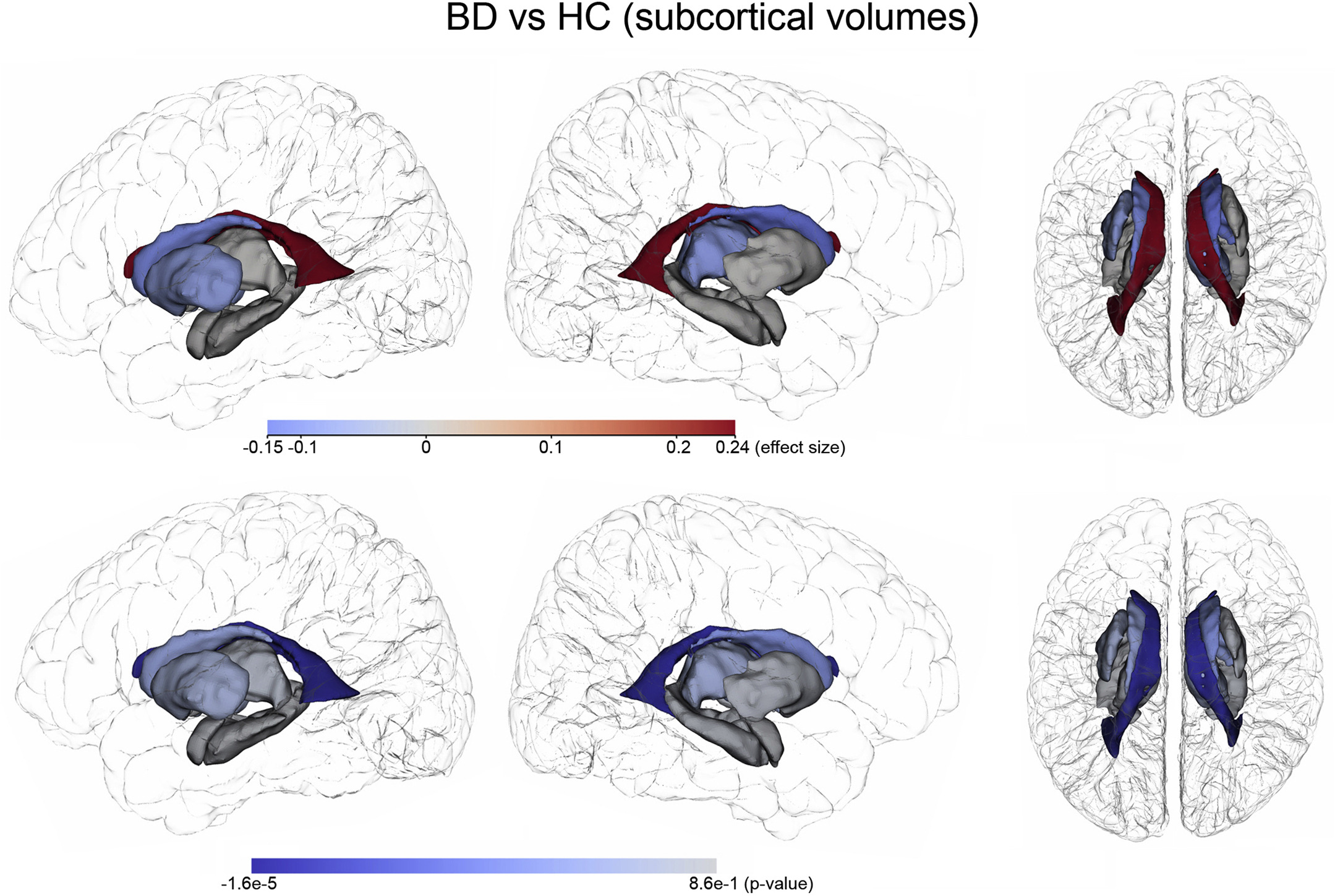
Subcortical volume findings between patients with bipolar disorder (BD) and healthy control (HC) subjects. Photo credit: USC Stevens INI
When it comes to mental health specifically, society at large still tends to disembody afflictions of the brain. Common phrasing such as “it’s all in your head” suggests a false separation between mind and body – between the structure of the brain and how it creates our thoughts, behaviors, and overall physical health. Yet, through advances in the field of neuroimaging, we are gaining a better understanding of how brain abnormalities lead to major psychiatric illnesses.
Bipolar disorder (BD) is a serious mental health disorder characterized by disruptive recurrences of mania and depression that may last for weeks, months or even years. Researchers have long suspected that BD may involve subtle abnormalities in brain structure that lead to the debilitating symptoms of the disorder. Small cross-sectional (single visit) brain imaging studies of people with BD have supported this, but interpreting data collected from a single snapshot in time can only reveal so much.
Now, a multi-center longitudinal study formed by the ENIGMA Bipolar Disorder Working Group (ENIGMA-BD) shows abnormal changes over time in the brains of people with BD. In 2012, ENIGMA-BD formed a large collaborative neuroimaging team combining existing research samples from around the world to advance understanding of the brain mechanisms underlying BD. In this latest study, USC researchers along with researchers from the University of Gothenburg, gathered magnetic resonance imaging (MRI) and detailed clinical data from 307 people with BD and from 925 healthy controls (HC) from 14 clinical sites worldwide. Participants were assessed at two occurrences, ranging from six months to nine years apart.
“The goal of the ENIGMA Bipolar Disorder Working Group is to bring researchers together from around the world so that we can combine existing data samples and really improve our power to detect the subtle brain alterations associated with this illness. While we have learned a great deal about how the brain is altered in bipolar disorder, very few large-scale studies have attempted to map the progressive neuroanatomical changes in this illness over time,” says Paul M. Thompson, PhD, of the Keck School’s USC Mark and Mary Stevens Neuroimaging and Informatics Institute (USC INI).
“In this first longitudinal study from our ENIGMA-BD team, and the largest study of its kind ever conducted, we map the subtle alterations in brain trajectories in those with BD over time and find that those who experience a greater number of manic episodes may show faster rates of frontal cortical thinning over time,” notes Christopher R. K. Ching, PhD, a postdoctoral fellow working with Thompson.
While this work provides important new clues about the structural efforts of BD on the brain over time, it also emphasizes the importance of longitudinal studies and large, diverse research cohorts. “This study exemplifies why the work at the USC INI is so essential,” says INI Director Arthur W. Toga, PhD. “Our innovative imaging and information technologies and our commitment to sharing data and resources allows us to be particularly supportive of large-scale studies. Our research does not exist in isolation, and we take great pride in what we have to share and gain from our peers in the scientific community.”
This research was supported by the National Institutes of Health and by many private and public funding agencies worldwide. The full paper can be access here.
07/27/2021
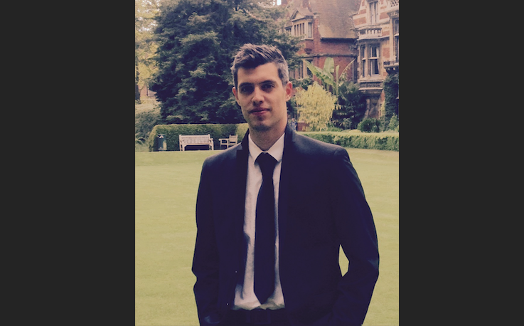
Join us in welcoming the newest member of the INI faculty, Ioannis Pappas, PhD, who conducts multimodal studies of patients with brain injury and stroke.
Brain imaging and computational models offer insights that may lead to recovery among patients who are comatose or in a persistent vegetative state. But studying such patients is a challenge. By observing individuals under general anesthesia—a more controlled loss of consciousness—Ioannis Pappas, PhD, can begin to understand what’s happening in the brain and build models to test in patients with brain injuries.
Dr. Pappas, who joined the USC Mark and Mary Stevens Neuroimaging and Informatics Institute (INI) as an assistant professor of research neurology in July, conducted that research during his doctoral studies in clinical neuroscience at the University of Cambridge in England. He used functional magnetic resonance imaging (MRI), as well as structural methods including diffusion tensor imaging (DTI), to study how neural connectivity changes in patients who are unconscious and whether those changes can predict recovery over the long term.

During his postdoctoral studies at the University of California Berkeley’s Helen Wills Neuroscience Institute, Dr. Pappas shifted his research focus to stroke recovery, with funding from the U.S. Veterans Administration. Using fMRI, DTI, blood flow imaging, and spectroscopy, he collected longitudinal data of veterans recovering from a stroke in an effort to understand the relationship between behavioral and neurological changes over time.
Because of the nature of that data—stroke patients have lesions in the brain that differ from one person to the next—a big part of Dr. Pappas’ research involves developing methods to standardize and facilitate the processing of MR images. Collecting data with multiple methods, such as combining standard structural brain images with arterial spin labeling, also helps create a more complete picture of stroke recovery. For example, Dr. Pappas found that the amount of residual blood flow in chronic stroke patients who have aphasia relates to their scores on language tests.
At the INI, Dr. Pappas will continue using multimodal methods to characterize clinical data. He is excited to draw on the institute’s vast collections of data and collaborate with its multidisciplinary team of researchers that includes engineers, cognitive neuroscientists, imaging experts, and more.
“We cannot have just one approach—we need multiple ways of collecting, analyzing, and interpreting neuroimaging data,” he said. “For that reason, interdisciplinary research is the way forward for tackling big programs of pathology and disease in the brain.”
Read more about Dr. Pappas’ findings on sedation and disorders of consciousness:
Brain network disintegration during sedation is mediated by the complexity of sparsely connected regions in NeuroImage
Consciousness-specific dynamic interactions of brain integration and functional diversity in Nature Communications
Read more about his research on methods development for studying stroke recovery:
Improved normalization of lesioned brains via cohort-specific templates in Human Brain Mapping
Poster presentation from the 2020 Society for the Neurobiology of Language conference: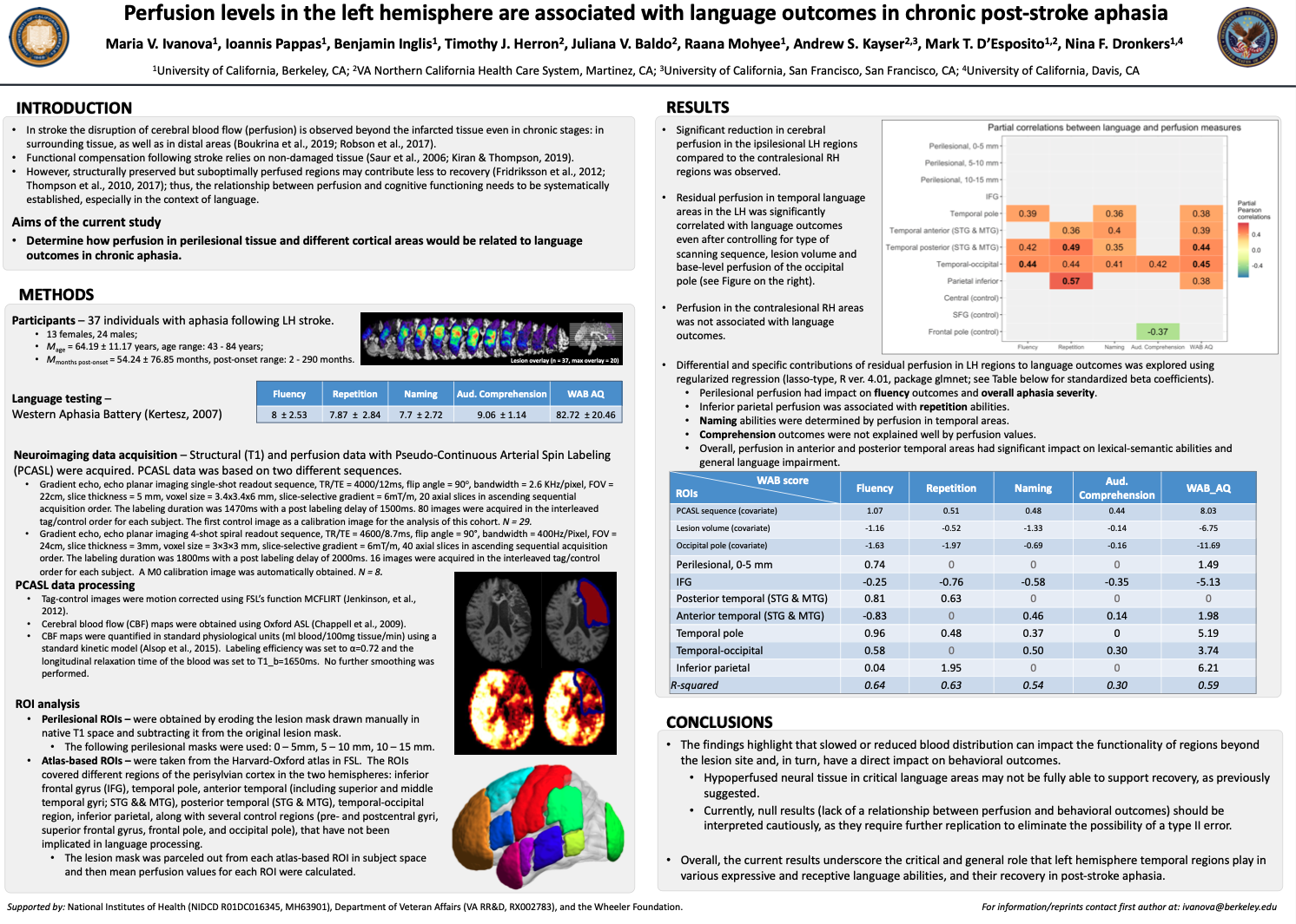
06/03/2021
Join us in welcoming the newest member of the INI faculty, Leon Aksman, PhD, who is applying insights from his background in engineering, neuroimaging, and financial modeling to examine the pathology of Alzheimer’s disease.
Dr. Aksman’s career path has been a bit unorthodox. After completing bachelor’s and master’s degrees in engineering, he worked in finance for several years. When he returned to academia to pursue a doctorate in neuroimaging, he applied his knowledge of quantitative modeling to one of the field’s most pressing questions: What causes Alzheimer’s disease (AD) and how does it progress over time?
After completing his doctorate at Kings College in London and postdoctoral training at University College London, Dr. Aksman joined the faculty at the USC Mark and Mary Stevens Neuroimaging and Informatics Institute last fall. Here, he is continuing his work applying statistics and machine learning to multimodal neuroimaging data, as well as cognitive and biofluid measures.

“Because there aren’t yet effective treatments for Alzheimer’s, what really drives me is to understand the disease better,” he said. “We can develop better treatments when we know more about the sequence and heterogeneity of pathologies in AD.”
For example, he used a mathematical model to analyze neuroimaging data from the Alzheimer’s Disease Neuroimaging Initiative (ADNI), including subjects in early, middle, and late stages of developing amyloid and tau pathologies. His model revealed two subtypes: one where amyloid pathology develops first and one where tau pathology develops first. This finding challenges the prevailing view among AD researchers that amyloid pathology always accumulates first.
“We got this result in a very data-driven way, but now we have to confirm it using both longitudinal and postmortem data,” Dr. Aksman said.
This research can help answer questions about whether AD manifests differently in certain patients, which can ultimately inform the development of targeted treatments.
Dr. Aksman is also analyzing data from Longitudinal Aging Study in India Diagnostic Assessment of Dementia (LASI-DAD), which includes data on adults in India who have developed dementia, with measures of literacy and urban/rural habitation. He will explore whether AD progression can be classified into various subtypes among this population.
“Most Western studies have not captured the full spectrum of socioeconomic variation in cognitive decline, which may mean that we have yet to explore the full heterogeneity of dementia,” he said.
09/24/2020
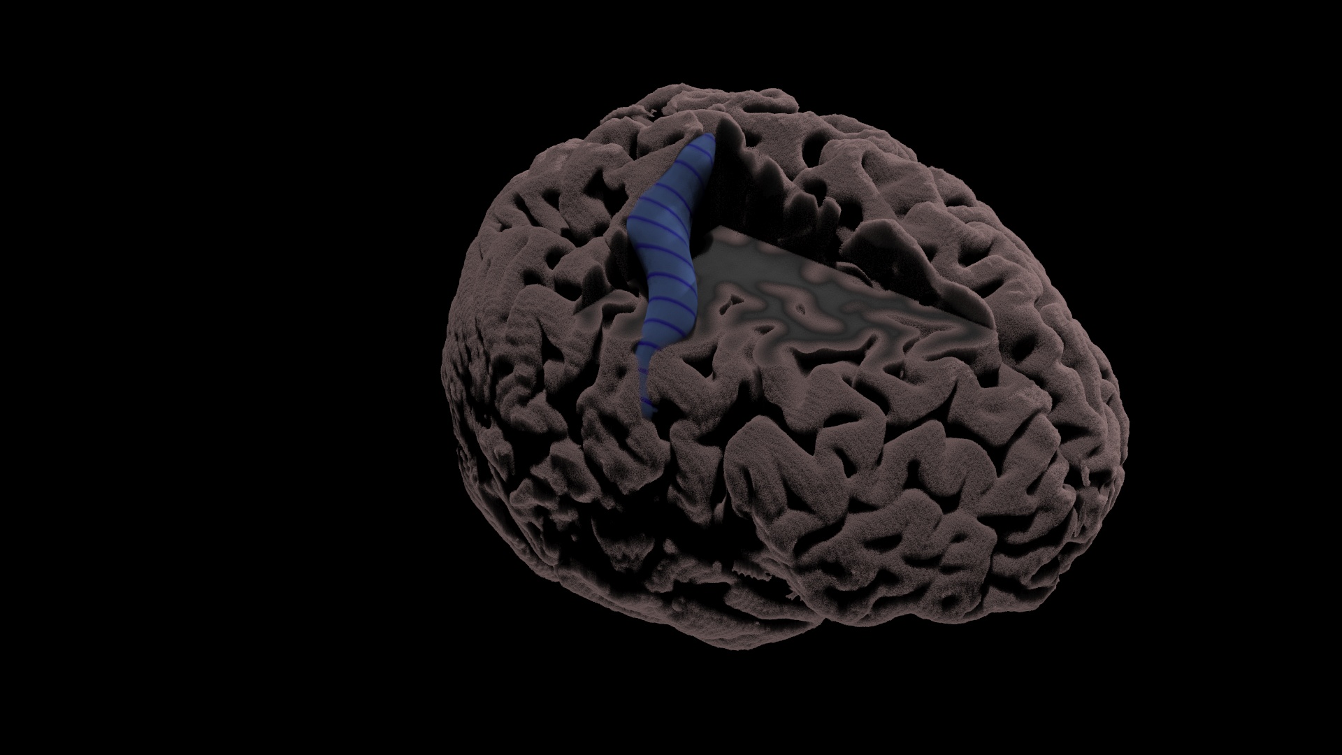
Neuroscientists around the world are working to better understand the genetic basis of brain structure and all of its complexities. A new study, published in the Nature journal Communications Biology in September and co-led by researchers at the USC Mark and Mary Stevens Neuroimaging and Informatics Institute (USC INI), analyzes cortical folding—the structural folds in the brain’s cortex—as a key piece of this puzzle. The research shows that cortical folding patterns are highly heritable—and that in some regions of the cortex, the genetic signature differs between the brain’s two hemispheres.

The team, led by Fabrizio Pizzagalli, PhD, who completed a postdoctoral fellowship at the USC INI and is now an assistant professor of applied physics in the Department of Neurosciences “Rita Levi Montalcini” at the University of Turin in Italy, performed a meta-analysis of more than 9,000 brain scans from MRI datasets around the world. The researchers measured the heritability of 61 sulci—grooves in the brain’s surface—in each hemisphere of the brain. Their findings suggest that analyzing cortical folding can provide key insights into the genetic factors that determine what each brain hemisphere controls.
"Now that we know which sulci can be robustly segmented, and we know their heritability, we can start to explore the processes underlying variation in human brain structure, beyond more traditional measures such as cortical thickness and surface area,” says Pizzagalli. “In particular, we can study the association of sulcal-based morphometry with diseases and the underlying genetic risk factors.”
Neda Jahanshad, PhD, associate professor of neurology at the USC INI’s Imaging Genetics Center, was Pizzagalli’s mentor and senior author of the study. Other contributors to the study included Josh Boyd, a graduate of the institute’s Master of Science in Neuroimaging and Informatics (NIIN) program.
09/22/2020
While neuroimaging is a valuable window into what happens inside the brain, it’s still an imprecise measure. Even after decades of technological advances, magnetic resonance imaging (MRI) scans still aren’t sharp enough to reveal what’s going on at the cellular level.
“By looking at brain scans, we don’t always know what mechanisms are driving a pattern of change in a given illness,” says Paul Thompson, PhD, professor of ophthalmology, neurology, psychiatry and the behavioral sciences, radiology, and engineering and associate director of the USC Mark and Mary Stevens Neuroimaging and Informatics Institute. “Researchers are left with a bit of a puzzle as to what those changes represent.”
But now, a revolutionary partnership between Thompson’s Enhancing Neuroimaging Genetics through Meta-Analysis (ENIGMA) consortium—a massive data-sharing effort between scientists in 45 countries—and researchers at the University of Toronto has led to a new method that can help pin down the mechanisms behind key psychiatric and developmental disorders. It’s a powerful breakthrough that bridges the gap between imaging and genetics research to identify the specific cells and molecules at play.
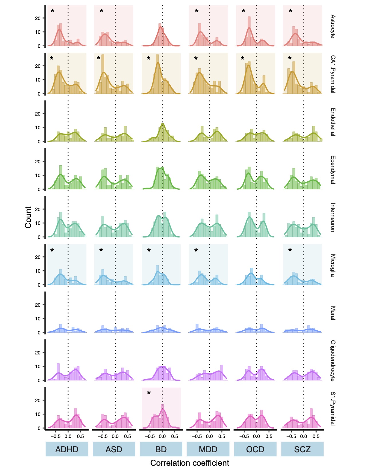
The team analyzed data on cortical thickness from more than 28,000 disease patients and healthy controls, ages 2 to 89, including individuals with schizophrenia, bipolar disorder, major depressive disorder, obsessive-compulsive disorder, autism spectrum disorder and attention-deficit/hyperactivity disorder. They identified a common molecular signature that spans all six disorders, publishing their findings in the journal JAMA Psychiatry in August.
The results can help pharmaceutical companies develop drug treatments that more directly address brain abnormalities, while the method developed by the team can be applied to other illnesses gain innumerable insights into the brain.
“This is a unique opportunity for science,” says Tomáš Paus, MD, PhD, professor at the University of Toronto and senior author of the paper. “ENIGMA has helped drive social innovation that allows us to move forward incredibly quickly—and to generate powerful new knowledge.”

LINKING IMAGING AND GENETICS
The new method developed by Paus and his team, known as “virtual histology,” uses atlases of gene expression to connect the dots between MRI scans and what’s happening in the brain at the cellular level. That’s possible because specific groups of genes mark certain types of cells in the brain.
For example, imaging data revealed that one cell type in particular—the pyramidal neuron—often underlies the MRI signature of the diseases studied.
“Regions that have more of these cells show a bigger difference in cortical thickness between disease cases and controls,” Paus says.
Building on this finding, Yash Patel, first author of the paper and a doctoral student in Paus’ lab, conducted a “co-expression analysis,” which identifies the genes involved in shaping those cells and plots their expression over time.
“We found two groups of genes responsible for controlling those pyramidal cells—one prenatal and one postnatal,” says Patel.
The prenatal genes control the development of axons and other infrastructure in the brain, while the postnatal genes are related to plasticity and neurotransmission. Those findings suggest that people who develop schizophrenia, bipolar disorder and the other brain diseases studied may experience common environmental influences after birth that triggers disease onset, Patel says.
By applying the virtual histology method to the massive datasets aggregated by the ENIGMA network, the study’s authors have begun to uncover the biological mechanism that links genetic vulnerability for a disorder to actual changes in the brain.
NEXT STEPS
Looking forward, Paus and his team plan to conduct a similar analysis on the brain’s surface area and to apply the virtual histology method to a series of other problems, including Alzheimer’s disease, Parkinson’s disease and brain trauma.
“We can only develop practical remedies for these conditions if we know which genes, molecules and pathways to home in on,” Thompson says. “This method provides that crucial bridge for information to travel between the genetics and imaging communities.”
08/24/2020
The USC Mark and Mary Stevens Neuroimaging and Informatics Institute (USC INI) of the Keck School of Medicine of USC has received accreditation from the American College of Radiology (ACR) for its 7Tesla Siemens Magnetom Terra magnetic resonance imaging (MRI) scanner.
Widely recognized as the “gold standard” in medical imaging, the ACR has assessed various imaging modalities, including MRI, computed tomography (CT) and ultrasound, since 1987. It uses a series of quality and safety checks to assess scanners and scanning facilities before granting accreditation. The INI’s scanner was the first 7T system to receive accreditation from the ACR for neuroimaging.
“ACR accreditation provides patients and referring physicians with the assurance that we are operating our state-of-the-art equipment in a manner that reflects our commitment to quality and safety,” says Vishal Patel, MD, PhD, assistant professor of radiology at the Keck School and medical director of the INI’s Center for Image Acquisition.
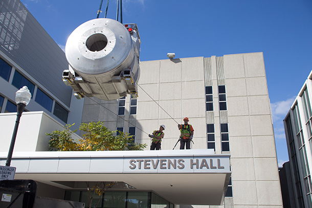
The institute’s 7T MRI scanner, the first of its kind in North America, was installed in 2017. It uses advanced technology and a powerful magnetic field to collect highly detailed images of the living brain that surpass the quality of standard MRI machines. This allows researchers to see neural structures and pathways in very high resolution and to more effectively study brain tumors, epilepsy and more.
In 2018, the scanner was also approved by the FDA for clinical use, establishing it as a safe and effective instrument for diagnosing and monitoring patients with neurological diseases such as multiple sclerosis and vascular dementia.
Now, the ACR’s stamp of approval further recognizes the INI’s proficiency in operating this unique diagnostic tool for both musculoskeletal imaging and neuroimaging.
“Uniting researchers and clinicians in their efforts to study and treat brain disease is essential to our mission as an institute,” says Arthur W. Toga, PhD, provost professor of ophthalmology, neurology, psychiatry and the behavioral sciences, radiology and engineering, the Ghada Irani Chair in Neuroscience at the Keck School, and director of the INI. “We maintain instruments like the 7T scanner so we can equip leading scientists with the most powerful tools available to fuel their endeavors.”
08/04/2020
After graduating from the INI’s Master of Science in Neuroimaging and Informatics (NIIN) program, Akul Sharma (’18) began working as a neuroimaging data analyst at the University of California Irvine’s Bio-behavioral Research on Adolescent Development (BRoAD) Lab. Akul is the primary MRI operator and imaging data analyst for two studies, both funded by the National Institutes of Mental Health. One involves studying the effects of childhood maltreatment on neurocircuitry in adolescent depression; the other explores ways to prevent adolescent risk-taking behaviors in African American youth.
Since graduation, Akul has published a number of abstracts, including presentations on adolescent depression and other topics at the annual meetings of the Organization for Human Brain Mapping, the American College of Neuropsychopharmacology and the American Academy of Child and Adolescent Psychiatry.
Looking forward, he plans to pursue a doctoral degree at the intersection of neuroimaging, psychiatry and informatics. Specifically, Akul hopes to use neuroimaging and neurocognitive measures to study emotional and cognitive dysfunction.
“The goal of my research is to increase our understanding of the existing neural models of mood disorders,” he says. “This can potentially improve clinical practice by enhancing biomarkers, treatment guidelines and algorithms.”
Below, Akul presented "White Matter Changes in Fronto-Limbic Pathways in Adolescent Depression" at the Society of Biological Psychiatry Annual Meeting. (Chicago, IL, May 2019)
10/29/2019
Since graduating from the INI’s Master of Science in Neuroimaging and Informatics (NIIN) program, Arvin Saremi (’17) hasn’t slowed down. He has coauthored four journal articles based on research he conducted with the INI’s Neda Jahanshad, PhD, during the master’s program, and is now a medical student at Case Western Reserve University, a top-25 ranked medical school for research.
“The NIIN program opened up many new doors for me—both in terms of opportunity and outlook,” Saremi says. “After exploring neuroimaging, I was able to enter medical school with a clear idea of my career interests from the beginning.”
*
During his year in the NIIN program, Saremi contributed to research at the INI’s Imaging Genetics Center, studying brain development in children with HIV, genetic markers associated with the volume of the hippocampus, and other topics. He recommends that future students seek out a faculty mentor at the institute and begin conducting research as soon as the program begins.
“The concepts taught in the program are incredibly complex. The best way to learn them is to actively apply them in research,” he says.
Because of the expertise he developed in diagnostic imaging during the NIIN program, Saremi was able to land a position in a neuroimaging lab just weeks after entering medical school. He says he’ll keep an open mind as he moves through the program but ultimately hopes to focus on neuroradiology.
Explore the publications Saremi coauthored during his time at USC:
Mapping abnormal subcortical neurodevelopment in a cohort of Thai children with HIV.
Structural Neuroimaging and Neuropsychologic Signatures in Children With Vertically Acquired HIV.
Novel genetic loci associated with hippocampal volume.
Keck School News,
06/27/2022
A USC-led team of researchers releases expanded data set of brain scans from stroke patients with more than four times the data in the hopes of speeding up large-scale stroke recovery research.
Keck School News,
06/14/2022
New findings from the largest study to date by an international group of neuroscience experts show significant reductions in grey matter in people with anorexia nervosa.
Keck School News,
06/07/2022
Research funded by the grant will capitalize on the development of biomarkers and advanced imaging by scientists at the Keck School of Medicine of USC to launch studies tracking changes in the blood-brain barrier, neurovascular function and cognition.
NPR,
04/26/2022
The INI's Paul M. Thompson is featured on All Things Considered.
Keck School News,
04/22/2022
The INI is proud to announce the appointment of Xingfeng Shao, PhD, to assistant professor of research radiology.
USC News,
04/06/2022
The findings could provide potential targets for new Alzheimer’s drugs.
Keck School News,
03/23/2022
Opened in 2016, the CIA is an MRI facility housing a Siemens Magnetom Prisma, a 3 Tesla MRI scanner, and a Siemens Magnetom 7T MRI scanner. These scanners include unparalleled innovations for neuroimaging.
Keck School News,
02/08/2022
As a result of a conference hosted by the INI, Brain Imaging and Behavior has published Special Issue: 2020 Pacific Rim New Horizons in Human Brain Imaging: Neuroimaging across the Lifespan.
Keck School News,
01/27/2022
USC’s recently launched Center for Neuronal Longevity brings together a multidisciplinary team to address unmet needs in vision loss and other neurological diseases
Keck School News,
01/20/2022
This serious mental health disorder comes with dramatic and sometimes unpredictable shifts in mood, energy and activity levels.
Keck School News,
01/12/2022
USC study suggests targeted outreach and better vaccine access may help reduce health disparities in vulnerable communities statewide.
Keck School News,
01/11/2022
“Our project aims to understand the connection between poor sleep leading to AD in African Americans. We will employ leading neuroimaging technologies at the USC INI to map the brain clearance system in individuals with and without sleep problems,” says Dr. Choupan.
Keck School News,
12/06/2021
INI Director Arthur Toga makes Clarivate’s Highly Cited 2021 list.
Keck School News,
12/02/2021
The INI is honored to have a section of the video, Ultrahigh Field MRI – The Value of Fine-Grained Examination, featured on the NIH Director’s Blog.
Keck School News,
11/22/2021
Danny JJ Wang Wang and his team are addressing the growing concern that radiation involved in CT scans can be unnecessarily harmful to patients.
Keck School News,
10/27/2021
Studies at USC’s Stevens Neuroimaging and Informatics Institute (INI) use mice to gain insight into brain structure and function to better understand brain disease and potential treatments.
Keck School News,
10/11/2021
The USC Mark and Mary Stevens Neuroimaging and Informatics Institute (USC Stevens INI) is one of three institutions partnering in a five-year, $31.7 million grant from the National Institutes of Health (NIH) to merge many types of Alzheimer’s disease data in an effort to stimulate new drug development.
Medical Xpress,
09/30/2021
A new USC study of more than 80 counties nationwide reveals the difference more or less restrictive policies may have had on the spread of COVID-19.
USC News,
09/21/2021
More than 90 federal grants totaling $92 million were awarded to USC researchers in 2020, underscoring the university’s status as one of the nation’s leading research centers as we acknowledge World Alzheimer’s Day 2021.
Keck School News,
09/10/2021
The National Institutes of Health is funding a global consortium of researchers from 20 countries to study how the disease develops and spreads.
USC Viterbi News,
07/14/2021
The USC+Amazon Center on Secure and Trusted Machine Learning, established in January 2021 to support fundamental research and development of new approaches to machine learning (ML) privacy, security, and trustworthiness, today announced it has selected five research projects for 2021-2022.
Physics World,
06/10/2021
A novel non-invasive neuroimaging technique can detect early-stage dysfunction of the blood–brain barrier (BBB) associated with small vessel disease (SVD), according to new research published in Alzheimer’s & Dementia. Cerebral SVD is the most common cause of vascular cognitive impairment, with many cases leading to dementia.
University of Kentucky,
05/07/2021
Collaborative research between the University of Kentucky and the University of Southern California (USC) suggests that a noninvasive neuroimaging technique may index early-stage blood-brain barrier (BBB) dysfunction associated with small vessel disease (SVD).
Trojan Family Magazine,
05/03/2021
When disease hides in the body, it takes some big ideas from scientists and doctors to illuminate it — and save lives.
Psychological Science Observer,
03/04/2021
Mapping the brain's unknown territories and vibrant connections
HSC News,
03/03/2021
It’s common knowledge that our surroundings affect our health — decades of research have linked things like air, water and soil quality to various measures of physical well-being. But much less is known about how the environment changes our brain. Now, a research team at the Keck School of Medicine of USC is pooling thousands of brain scans to better understand how aspects of our physical space — including air pollution, noise and green space — alter brain structure, influence our behavior and impact our risk for various developmental, neurodegenerative and psychiatric problems.
LONI,
02/18/2021
A stroke occurs when a blocked artery can no longer supply blood to part of the brain. But the mechanism differs between thrombotic strokes, embolic strokes, thromboembolic strokes, and brain hemorrhages. Our latest scientific visualization describes various types of strokes and illustrates what’s going on in the brain when they occur.
Nature Reviews Neurology,
02/02/2021
Two new studies, published in Nature Medicine and Movement Disorders, have shown that people with Parkinson disease (PD) exhibit alterations in brain lymphatic and glymphatic drainage systems. The findings suggest that dysfunction of brain drainage pathways represents both an early biomarker and a potential therapeutic target for PD.
HSC News,
01/15/2021
Nearly 38 million people around the world are living with HIV, which, with access to treatment, has become a lifelong chronic condition. Understanding how infection changes the brain, especially in the context of aging, is increasingly important for improving both treatment and quality of life.
HSC News,
01/04/2021
Until recently, scientists didn’t know much about perivascular spaces (PVS), fluid-filled regions in the brain involved in clearing waste and toxins — mainly because it’s tough to get a clear look at them using neuroimaging. But as technology evolves, researchers are finding that PVS may play a central role in brain changes associated with aging, including in neurodegenerative disorders such as Alzheimer’s disease.
HSC News,
12/07/2020
Ryan P. Cabeen, PhD, a postdoctoral researcher in the USC Mark and Mary Stevens Neuroimaging and Informatics Institute (USC Stevens INI) at the Keck School of Medicine of USC, has received an imaging technology grant from the Chan Zuckerberg Initiative (CZI), a philanthropy led by Priscilla Chan, MD, and her husband, Facebook founder Mark Zuckerberg.
HSC News,
10/22/2020
Researchers, led by a team at USC, will use artificial intelligence to study tens of thousands of brain images and whole genome sequences.
HSC News,
10/21/2020
Decreased blood flow is linked to more severe tau pathology in regions of the brain affected by Alzheimer’s disease.
US News & World Report,
10/19/2020
Offering fresh insight into the deep-seated roots of dementia, new research finds that diminished blood flow to the brain is tied to buildup a protein long associated with Alzheimer's disease.
HSC News,
09/03/2020
Researchers based at the USC Mark and Mary Stevens Neuroimaging and Informatics Institute have conducted the largest-ever diffusion imaging study of 22q11.2 deletion syndrome (22q11DS). The genetic disorder, caused by a small segment of missing DNA on chromosome 22, is the strongest known genetic risk factor for schizophrenia.
HSC News,
08/26/2020
For patients with tumors, strokes, cerebrovascular disease and other conditions, computed tomography (CT) scans can be lifesaving. But there’s a growing concern that the radiation involved is unnecessarily harmful to patients.
HSC News,
08/19/2020
Clinicians and researchers around the world are racing to collect data in order to better understand COVID-19 — including risk factors for severe cases, treatment strategies and the viability of various vaccines. But a vital element missing has been a wide-scale database for sharing information as research progresses.
USC Health,
08/11/2020
Clinicians need to act quickly to administer effective treatment when a patient has an acute ischemic stroke — the most common type — which occurs when a clot blocks blood flow through an artery in the brain.
USC News,
08/05/2020
USC’s Sook-Lei Liew, Judy Pa and James M. Finley are researching the benefits of virtual reality for patients with Alzheimer’s disease, Parkinson’s disease or stroke.
BioSpace,
07/31/2020
A multi-omics, multi-algorithm approach will unleash a new wave of understanding around Alzheimer’s disease and diagnosis, that in time, will make precision diagnostics practical, speakers predicted at the Alzheimer’s Association International Conference 2020 virtual event, held July 27-31.
HSC News,
07/14/2020
The Donald E. and Delia B. Baxter Foundation is supporting innovative biomedical research at the Keck School of Medicine of USC by granting $100,000 awards to two assistant professors.
HSC News,
06/02/2020
Researchers at the USC Mark and Mary Stevens Neuroimaging and Informatics Institute at the Keck School of Medicine of USC are forging ahead with their research programs.
USC News,
04/29/2020
Although scientists have long known APOE4 is a leading risk factor for Alzheimer’s disease, they were unsure how exactly it drives a decline in memory. USC researchers now believe they have an answer.
HSC News,
02/13/2020
The key to a better understanding of schizophrenia may exist in a genetic disorder so rare that researchers haven’t been able to conduct an adequate study — until now.
Inverse,
02/02/2020
In a new study led by Dr. Arthur W. Toga, director of the INI, researchers analyzed MRI scans from more than 11,000 people and found that the brains of those who smoke or drink frequently tend to age faster.
Larry King Now,
01/20/2020
Alzheimer’s is one of the most common diseases affecting older generations in America.In fact, a person is diagnosed with Alzheimer’s disease every 65 seconds. Larry King sits down with a panel of experts to explore the research being done and preventative measures to stop early onset.
HSC News,
01/10/2020
Researchers at the USC Mark and Mary Stevens Neuroimaging and Informatics Institute are working on safer ways to scan the brains of stroke patients in assessing damage.
HSC News,
12/02/2019
This fall, the USC Mark and Mary Stevens Neuroimaging and Informatics Institute (INI) received a $2 million competitive renewal from the Alzheimer’s Association to continue expanding its pioneering data-sharing platform, the Global Alzheimer’s Association Interactive Network (GAAIN).
HSC News,
12/01/2019
Paul M. Thompson, PhD, has received a Zenith Fellows research grant from the Alzheimer’s Association, one of the most prestigious awards in Alzheimer’s and dementia research.
HSC News,
11/01/2019
Most people know that problems with memory and cognition are an early hallmark of Alzheimer’s disease, but fewer are aware that in up to 88% of patients, neuropsychiatric issues like depression, anxiety and apathy are also among the first symptoms of the disease.
Annenberg Media,
10/29/2019
The app, created by a USC professor, has major implications for the medical field.
Spectrum,
10/23/2019
They say a picture is worth a thousand words. But what about a figure in a scientific paper?
INI,
10/21/2019
Learn about some of our latest findings, view our new scientific data visualizations & more in our interactive fall newsletter.
USC News,
10/17/2019
INI scientists have launched a smartphone app that uses augmented reality to visualize scientific data via 3D models and video.
National Institute on Aging,
10/16/2019
Brain imaging data will be stored at the Laboratory of Neuro Imaging at the University of Southern California in Los Angeles.
LONI Blog
10/14/2019
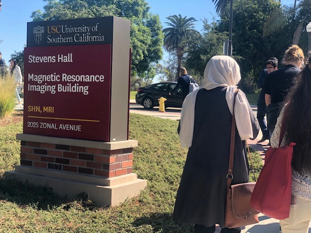
For the first time, the INI has convened the research and clinical imaging communities of Southern California for an impactful annual workshop on high- and low-field Magnetic Resonance Imaging technologies. On October 12, the group convened on the USC Health Sciences Campus for a day of tours, tutorials and presentations that connected researchers from across the region and stimulated new lines of inquiry as well as novel research partnerships.
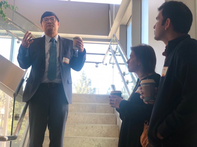
Danny JJ Wang, PhD, professor of neurology, director of imaging technology of innovation at INI and co-organizer of the workshop, started the day by welcoming guests to the institute and sharing a brief history of the INI and the Laboratory of Neuro Imaging Resource (LONIR), a platform for collaborative research in the neurosciences. Later, Wang led a tour of the institute and its Center for Image Acquisition (CIA), which houses both an ultra-high-field (UHF) 7-Tesla Siemens Terra MRI system and a 3-Tesla Siemens Prisma MRI system.
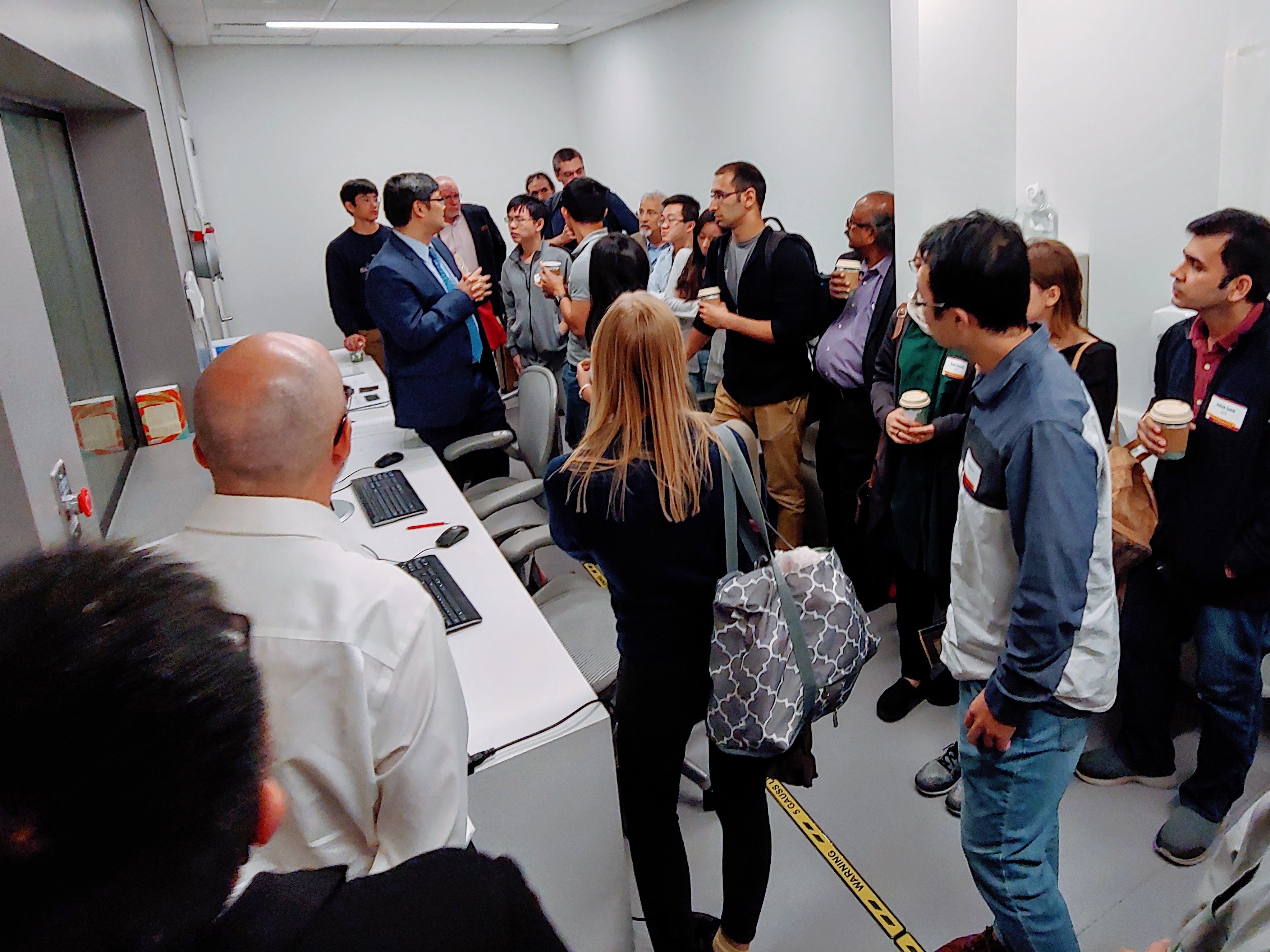
Presentations throughout the day included an in-depth discussion of high-performance-low-field (HPLF) MRI at USC by workshop co-organizer Krishna Nayak, PhD, a professor of electrical engineering at the Viterbi School of Engineering with joint appointments in radiology and biomedical engineering. Other topics, including clinical and research applications of UHF MRI, vascular imaging with UHF MRI, UHF coil developments, cardiac intervention and lung imaging with HPLF MRI, and more were also featured.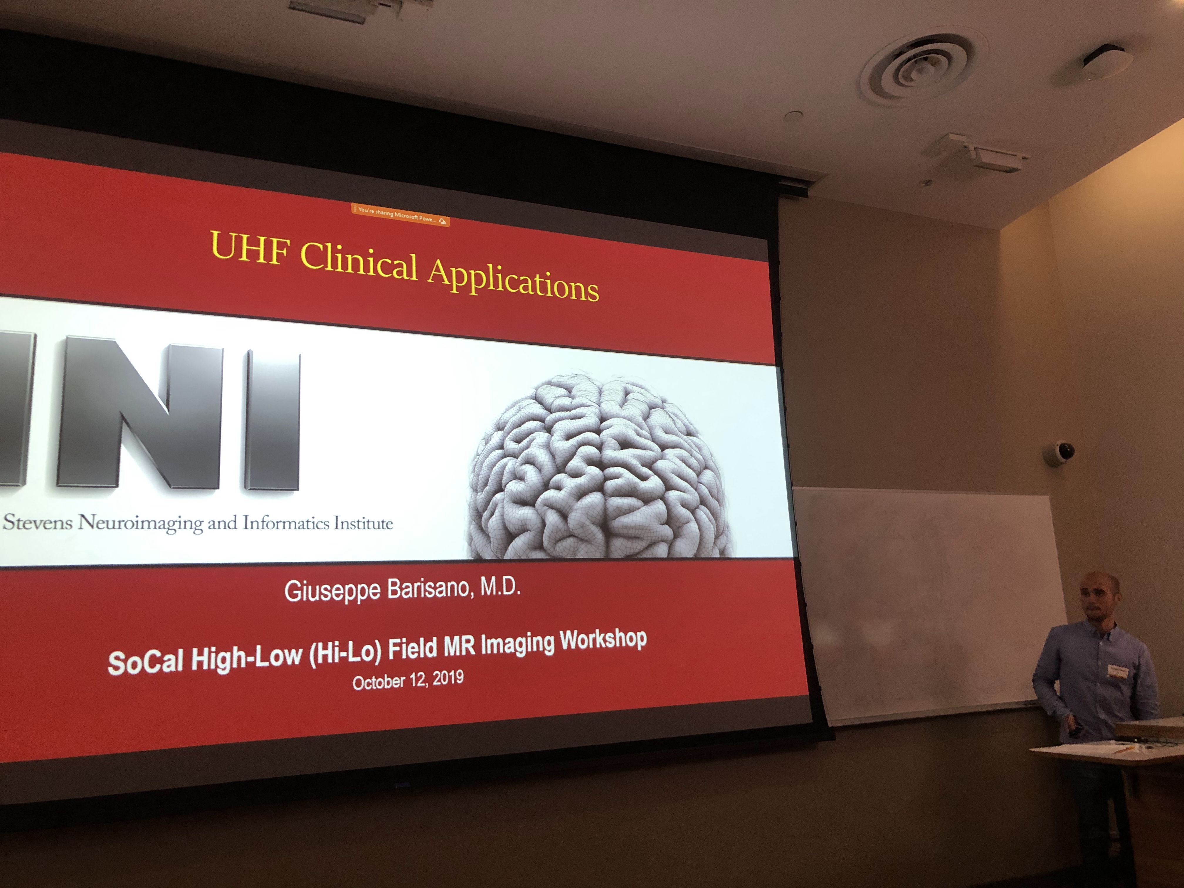
The event was sponsored by the Laboratory of Neuro Imaging Resource, the Center for Image Acquisition, and Siemens Healthineers.
VOA News,
09/07/2019
Many adult children are painfully seeing their parents experience cognitive decline and symptoms of dementia. What if virtual reality, or VR, can help prevent or delay the onset of cognitive decline?
GoldieBlox,
08/29/2019
Dr. Liew partnered with children's media company GoldieBlox for their new YouTube series, Fast Forward Girls.
HSC News,
08/28/2019
Meredith Braskie, PhD, assistant professor of neurology at the Keck School of Medicine of USC, has been named director of education for the USC Mark and Mary Stevens Neuroimaging and Informatics Institute (INI) effective Aug. 1.
MD Mag,
08/23/2019
A massive new dataset of RNA sequencing data is now included in the Parkinson’s Progression Markers Initiative (PPMI), a database which involves collecting clinical, biological, and imaging data from 1400 individuals over at least 5 years.
INI,
08/21/2019
The Southern California (SoCal) High Low (Hi-Lo) Field Workshop is an annual workshop that will help researchers maximize the unique resources and research programs on the ultra-high-field (UHF) 7T Terra system at the Stevens Neuroimaging and Informatics Institute and the high performance low field (HPLF) ~0.5T system at the Ming Hsieh Department of Electrical and Computer Engineering of the USC Viterbi School of Engineering. The goal is to engage the research and clinical imaging communities of Southern California, build a user base and stimulate new ideas.
HSC News,
08/21/2019
A team of experts at the INI, in collaboration with The Michael J. Fox Foundation for Parkinson’s Research (MJFF), has just added a crucial new element – RNA sequencing data — to its robust study of Parkinson’s disease.
NIH Director's Blog,
08/20/2019
One of LONI's animations was featured on the NIH Director's blog on August 20.
HSC News,
08/16/2019
On July 1, Vishal Patel, MD, PhD, joined the Mark and Mary Stevens Neuroimaging and Informatics Institute (INI) at the Keck School of Medicine of USC as an assistant professor of radiology.
AlzForum,
07/25/2019
Scientists are becoming more nuanced in how they use amyloid scans—not just to detect the presence of Alzheimer’s pathology, but also to pinpoint disease stage.
Chan Division News,
07/24/2019
INI's Sook-Lew Liew is showing young women what's possible in STEM.
LONI Blog
07/19/2019
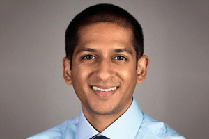
On July 1, Vishal Patel, MD, PhD, joined the INI as an assistant professor of radiology. Patel, who earned his doctoral degree in biomedical engineering, completed a residency in diagnostic radiology at the University of California, Los Angeles and a fellowship in neuroradiology at USC.
Patel’s research interests include MRI, diffusion imaging, tractography and machine learning. At the INI, he plans to apply his knowledge of computational MRI and artificial intelligence (AI) to improve diagnostic techniques for neuroradiologists.
“The goal is to establish USC, and in particular the INI, as a site where AI methods and techniques are developed, validated and made available for use to the greater neuroimaging community, including both researchers and clinicians,” Patel says.
AI holds promise for a number of clinical challenges. For instance, an MRI scanner may register images of very small brain lesions that the naked eye cannot detect, but researchers like Patel can build software that identifies subtle abnormalities present in the underlying imaging data. Ultimately, this technology can be used to detect tumors and classify them by cell-type and pathological grade, providing a precise and non-invasive way to diagnose brain cancer.
Patel’s research at USC will build on his previous work applying machine learning concepts to tumor identification and other clinical challenges. But now, he has a powerful new tool: the INI’s ultra-high field 7Tesla MRI scanner, which received FDA approval for clinical scanning last fall.
“The INI is proud to welcome Vishal to our team,” says Arthur W. Toga, PhD, provost professor at USC and director of the INI and the Laboratory of Neuro Imaging (LONI). “Applying his expertise in neuroradiology to our high-end clinical imaging system will undoubtedly lead to meaningful new discoveries.”
Toga and Patel have collaborated in the past and look forward to resuming their research together on a new clinical imaging study of patients with multiple sclerosis (MS).
“LONI was a phenomenal place during my graduate training and it continues to become more and more impressive, especially with the addition of clinical high-field imaging capabilities,” says Patel. “I’m excited to come on board and get to work in what I know to be an excellent environment for research.”
USC News,
07/15/2019
Understanding the brain structure of transgender people could help tailor care and support, says recent USC grad and neuroscientist Jonathan Vanhoecke.
USC News,
07/11/2019
USC research into Alzheimer’s — which will be on display at the upcoming Alzheimer’s Association International Conference — has uncovered much about the disease, including establishing a link between cardiovascular health and a fully functioning brain.
LONI,
07/03/2019
Check out this video of microstructure and connectivity in the brain, created by LONI's Jim Stanis using data collected by Ryan Cabeen and Arthur W. Toga.
USC Division of Biokinesiology and Physical Therapy,
06/20/2019
The interdisciplinary endeavor aims to establish USC as a world leader in the use of virtual technologies for healthcare.
HSC News,
06/11/2019
Shaina Sta. Cruz, a student researcher at the USC Mark and Mary Stevens Neuroimaging and Informatics Institute, authored her first publication in the journal Neuroinformatics.
HSC News,
05/08/2019
Spotlight on Jonathan Vanhoecke, Keck School of Medicine of USC
HSC News,
05/06/2019
Spotlight on Elizabeth Haddad, Keck School of Medicine of USC
INI,
04/22/2019
This spring, researchers at the USC Mark and Mary Stevens Neuroimaging and Informatics Institute have published new studies exploring Alzheimer’s disease, connectivity in the mammalian brain and new noninvasive imaging techniques.
LONI Blog
04/15/2019
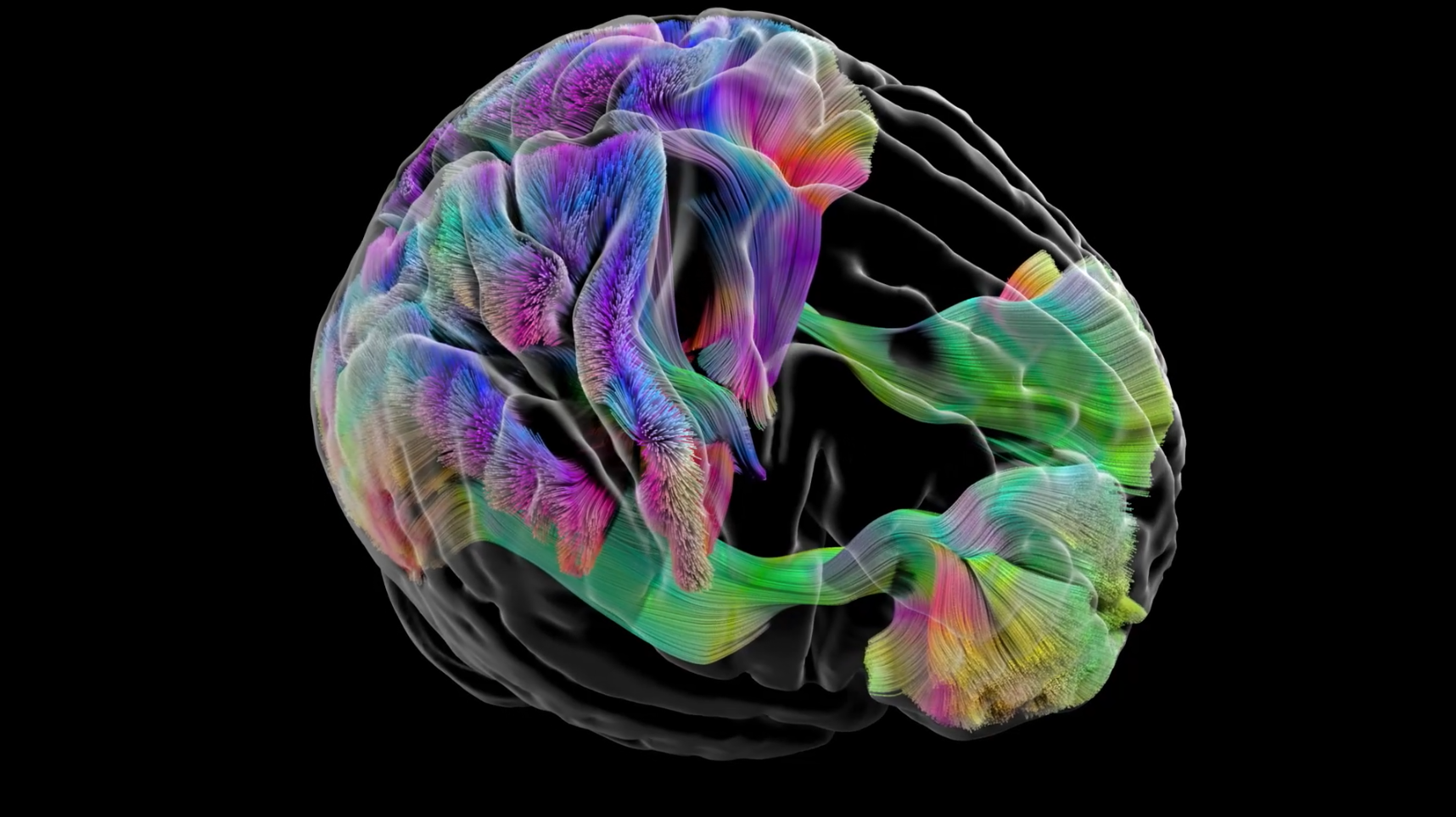
LONI's medical animator, Jim Stanis, won second place in the NIH BRAIN Initiative's 2019 video contest! His animation depicts 3D diffusion tractography, a modeling technique that maps connections in the brain.
HSC News,
03/21/2019
After months of hard work, dozens of students from Francisco Bravo Medical Magnet High School presented their original research projects at the annual Bravo-USC Science and Engineering Fair on March 4 to a panel of judges from USC, UCLA and other institutions.
USC News,
03/04/2019
INI master's student and radiation oncology specialist Faisal Rashid recently became the LAPD’s first Bangladeshi sworn officer through its reserve program
LONI Blog
02/14/2019
The blood brain barrier lines the vessels of the circulatory system in the brain, preventing harmful substances from crossing. This animation explains the blood brain barrier’s anatomy at a micro level—and what happens when it starts to break down.https://www.youtube.com/watch?v=vZCfTCF4ABo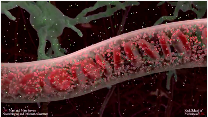
The INI is home to an interdisciplinary group of researchers, programmers and visualization specialists capable of producing sophisticated scientific visualizations. Videos like these help researchers better comprehend and communicate complex biological processes.
LONI Blog
01/29/2019

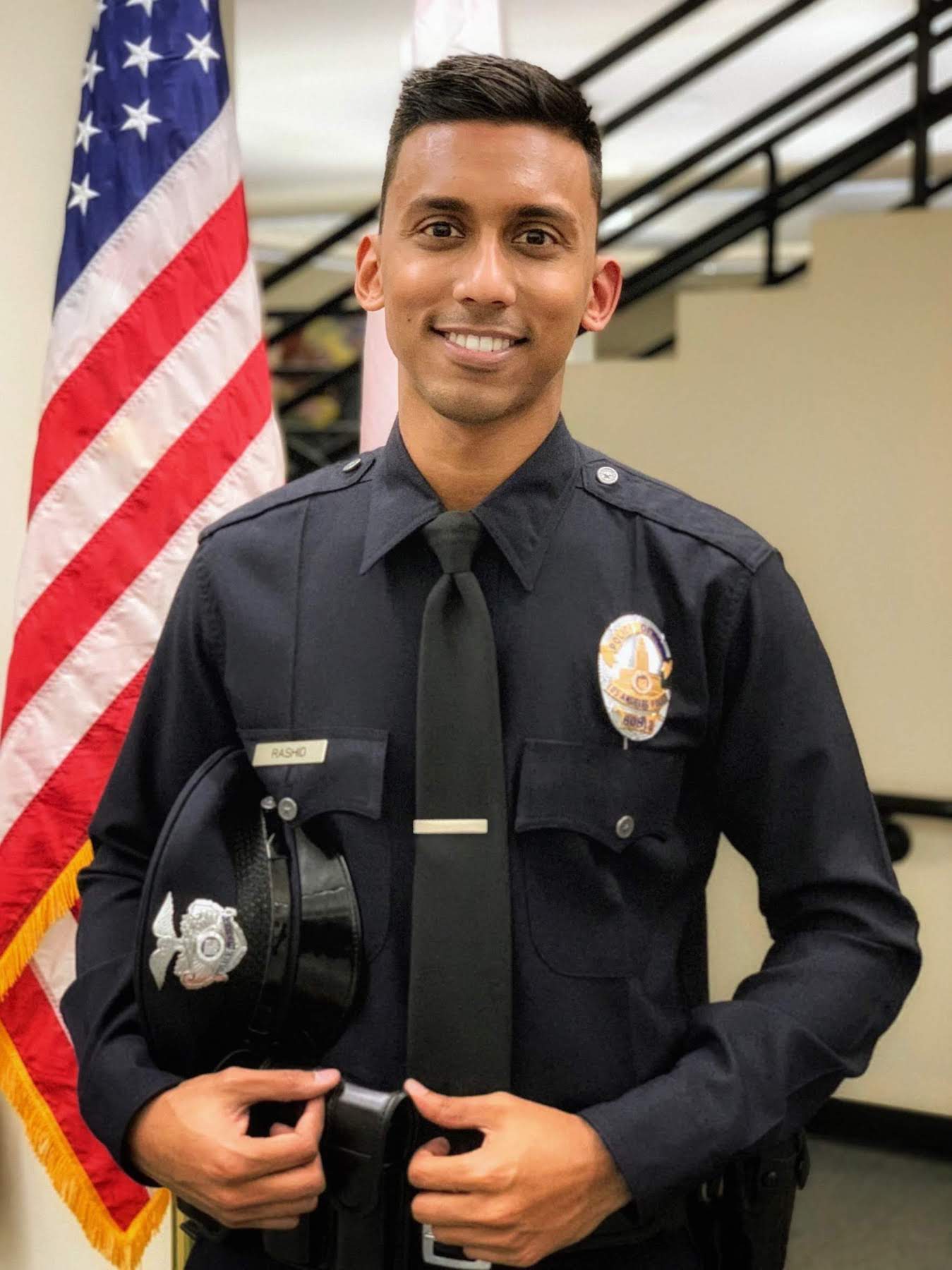
Faisal Rashid, a student in the INI’s Neuroimaging and Informatics master’s program, was sworn into the Los Angeles Police Department on January 4 as a reserve officer after nearly two years of interviews, background investigations, polygraph tests, extensive training, and medical, physical fitness and psychological examinations.
Rashid was inspired to join the academy while working as a project assistant in the lab of Paul Thompson, PhD, INI’s associate director. After studying how addiction, depression, post-traumatic stress disorder and other conditions affect brain health, Rashid says he began to understand how such issues contribute to crime and homelessness.
“Law enforcement officers are typically first responders to emergencies that involve individuals with mental health problems, trauma, or troubled adolescents. Sometimes, the right interaction with the right people can change the trajectory of someone’s life for the better," Rashid says, adding that it’s always been a priority for him to give back to Los Angeles, where he was born and raised.
He is currently assigned to the LAPD's Counter Terrorism and Special Operations Bureau. Moving forward, he hopes to study the relationship between mental health, trauma in childhood and adolescence, and crime.
Viterbi News,
01/28/2019
USC researchers found “hidden factors” in medical data that could improve Alzheimer’s disease prediction and lead to better outcomes.
US News & World Report,
01/22/2019
Leaky blood vessels in the brain may be an early sign of Alzheimer's disease, researchers say.
USC News,
01/15/2019
Brain changes associated with leaky capillaries suggest new, potential drug targets as well as a way to diagnose the disease sooner.
ENIGMA,
01/14/2019
The ENIGMA Consortium's new international initiative aims to better understand variables that affect brain aging and progression to dementia in diverse settings.
NIH Director's Blog,
01/10/2019
INI's recent Nature Neuroscience hippocampus study is featured on the NIH Director's Blog.
LONI Blog
12/18/2018

On December 14, INI held its holiday party and 2nd annual Winter Games competition. While faculty and staff enjoyed festive food, treats and cocktails, six teams entered in a fierce battle for first place in the highly anticipated Games.
Event One was a Pseudoscience Fair, in which teams created scientific posters to prove unscientific concepts, including birthday astrology, phrenology and crystal healing. Some teams conducted live “experiments” or used sweets, props and pandering to win over the judges.

Event Two required each team member to taste and name one of Bertie Bott’s Every Flavor Beans (a mainstay of the Harry Potter universe). Participants correctly identitifed a range of bean flavors, including everything from candyfloss and cinnamon to soap, earwax and vomit.
For Event Three, each team used a variety of building materials (including tinker toys, pipe cleaners, egg cartons and cardboard) to construct a scene related to the institute. Several teams crafted versions of the INI’s 7T MRI scanner. Teams also earned bonus points for wearing nametags adorned with photos of themselves dating back more than 10 years.

The judges deliberated carefully and ultimately named Carinna, Avnish, Jeana, Zach and Kaelan winners for their superb presentation on “The Effects of Phrenology on Collaborative Efforts in a Neuroscience Environment” and their outstanding jelly-bean tasting and construction skills.

LONI Blog,
12/06/2018
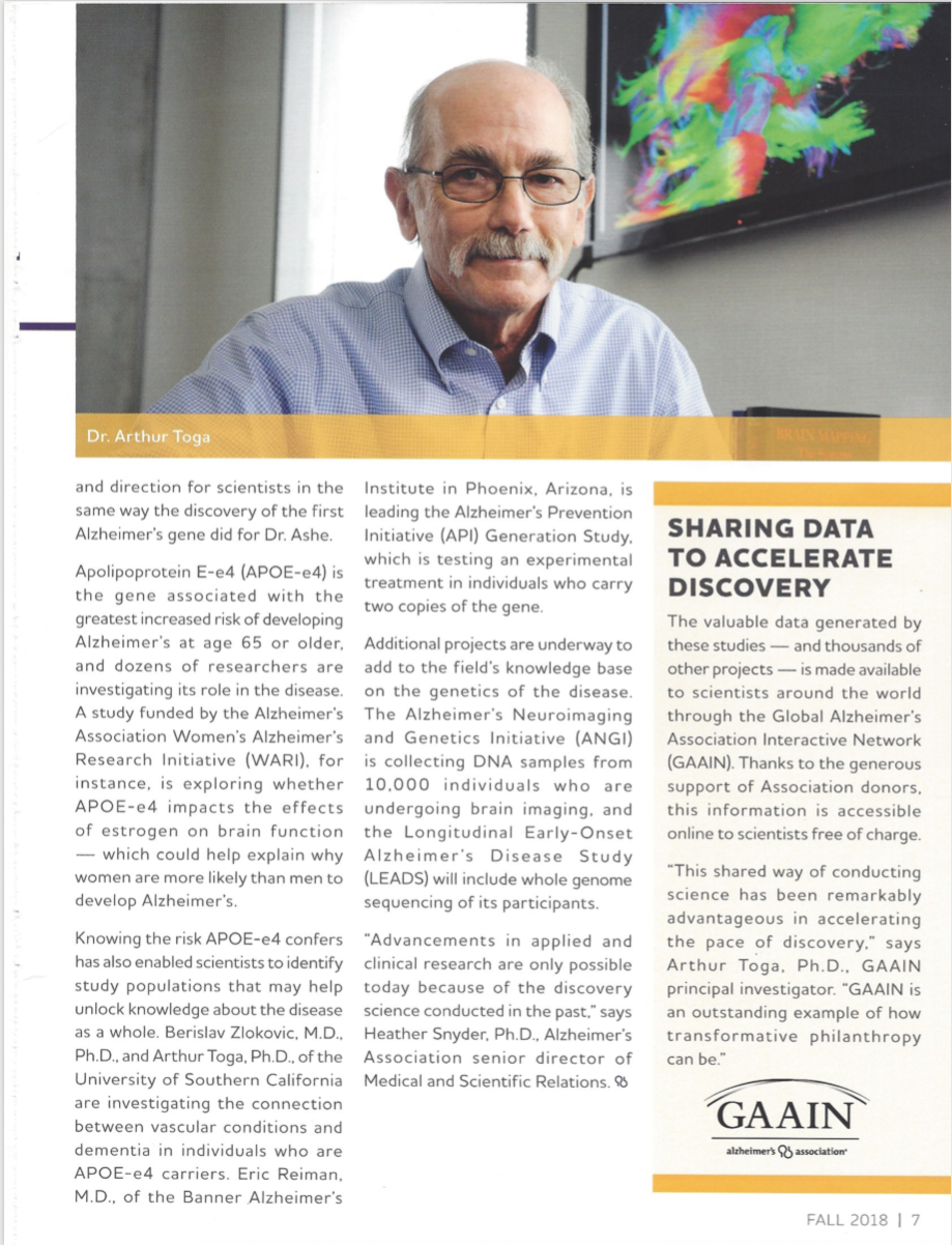
ASPIRE, the Alzheimer's Association philanthropy magazine, featured our Global Alzheimer's Association Interactive Network (GAAIN) and its PI, Arthur W. Toga. The network facilitates data exploration and discovery for Alzheimer's and aging researchers while allowing data owners to maintain control over their material. Read the text of the article below.
Clarivate Analytics,
11/27/2018
Paul M. Thompson, the associate director of INI, has been named one of Clarivate Analytics' 2018 Highly Cited Researchers.
Lab Animal,
11/19/2018
Not long after moving to southern California to start his postdoc with Hongwei Dong, Mike Bienkowski found himself at a meeting listening to neuroscientists debate the divisions of the hippocampus.
Aunt Minnie,
11/14/2018
Currently, three institutions in the U.S. have begun using 7-tesla magnets for clinical applications, but mostly for neurological and musculoskeletal conditions. The challenge now is showing how much more 7-tesla MRI can contribute to patient care.
USC News,
11/09/2018
Paul Thompson and his team just launched the ENIGMA World Aging Center, which will use the consortium’s network and a $682,000 grant from the National Institute on Aging to perform a global study of brain aging. The collaboration will help pinpoint the factors that contribute to healthy aging versus those that increase our risk for Alzheimer’s or Parkinson’s disease.
USC News,
10/30/2018
On October 24, the ultra-high-field 7T Terra magnetic resonance imaging (MRI) scanner at the USC Mark and Mary Stevens Neuroimaging and Informatics Institute (INI) of the Keck School of Medicine of USC received FDA approval for clinical use, opening up new avenues of care for patients with Alzheimer’s disease, multiple sclerosis and other diseases that affect the brain.
AlzForum,
10/19/2018
Now, the first neuronal-connectivity study to zoom in to the cellular level reports that the mouse hippocampus can in fact be divided into 22 discrete subregions. To find them, researchers led by Michael Bienkowski and Hong-Wei Dong at the University of Southern California, Los Angeles, compared gene expression and connectivity cell by cell.
LONI Blog
10/18/2018

The Virtual Brain Segmenter (VBS), a virtual reality tool for processing brain scan images, has been named a finalist for the 2018 Auggie Breakthrough Awards, which honor innovative academic-industry collaborations in virtual and augmented reality.
VBS was developed by INI’s Dominique Duncan, PhD, Tyler Ard, PhD, Arthur W. Toga, PhD and the industry group RareFaction Interactive to transform a tedious step in the scientific process into an immersive experience. After collecting MRI data, researchers typically correct errors in scan images by hand, but VBS allows them to speed up the process with a VR headset, joystick and larger-than-life images of the brain.
In an experimental trial, published in the Journal of Digital Imaging in July, participants finished a correction task 68 seconds faster using VBS compared with the traditional method, a highly significant time savings considering the task rarely took more than three minutes to complete.
Now, Dr. Duncan is traveling in Munich, Germany to represent INI at the Auggie Breakthrough Awards ceremony, where winners will be chosen in several categories, including Most Innovative Breakthrough, Most Impactful Breakthrough and Best in Show. Fellow finalists include representatives from NASA’s Jet Propulsion Laboratory, Stanford, the Mayo Clinic and international collaborators from Finland, Australia and beyond. Projects span the health sciences, communications, education, resource management and gaming.
Read more about the finalists in the official press release
Learn more about VBS from USC News
USC News,
10/08/2018
Researchers show structures, nerve connections and functions in vivid detail as part of the most detailed atlas yet of a person’s memory ban
USC News,
10/04/2018
Peer support group started by students for students lifts those who want a conversation with someone their own age.
HSC News,
09/26/2018
In fiscal year 2017-2018, the USC Mark and Mary Stevens Neuroimaging and Informatics Institute (INI) at the Keck School of Medicine of USC received more than $30.5 million in active research funding.
Discover Magazine,
09/01/2018
A new preprint called “A systematic bias in DTI findings” could prove worrying for many neuroscientists. In the article, authors Farshid Sepehrband and colleagues of the University of Southern California argue that commonly-used measures of the brain’s white matter integrity may be flawed.
LONI Blog
08/31/2018
Earlier this month, six undergraduate students from California State University, Fullerton presented their summer research results to a room full of neuroscientists at the Mary and Mark Stevens Neuroimaging and Informatics Institute (INI). The collaboration is part of CSUF’s Big Data Discovery and Diversity through Research Education Advancement and Partnerships (BD3 REAP) program, which trains underrepresented students in big data science.
“The program exposes these young investigators to what modern science is about these days, which is the collection of ever larger data sets,” said Jack Van Horn, INI’s director of education and leader of the collaboration. “They’re learning about the challenges of big data, and how they can manage, model, and understand it.”
Building on several semesters studying data science and analytics at CSUF, each student selected a big data neuroimaging project to pursue this summer with the guidance of INI faculty members and postdoctoral researchers.
For their presentations, the students covered methods, results and next steps. Each created a slideshow and was encouraged to delve into the technical aspects of research performed. Focus areas included the Epilepsy Bioinformatics Study for Antiepileptogenic Therapy (EpiBioS4Rx), using gene expression to build an atlas of the hippocampus and the link between functional brain connectivity and familial Alzheimer’s disease.
The students received mentorship from INI faculty, including program director Jack Van Horn, Kay Jann, Tyler Ard, Dominique Duncan, Lirong Yan and Meredith Braskie.
“This has been a great opportunity for INI to expand its commitment to training the next generation of leaders,” said Arthur W. Toga, provost professor at USC and director of INI. “We were impressed by the students’ work and look forward to continuing our outreach efforts with CSU Fullerton.”

Alz Forum,
08/23/2018
Two-thirds of AD patients are women, leading to the commonly held assumption that women run a higher risk of dementia. In Chicago, Arthur Toga of the University of Southern California, Los Angeles, challenged that view with data gleaned from a meta-analysis of 58,000 participants in 27 AD studies.
HSC News,
08/23/2018
USC offers numerous institutional training opportunities for postdocs, including parallel lines of research on aging and Alzheimer’s disease. But the new project, “Training for the Multiscale and Multimodal Analysis of Biomarkers in Alzheimer’s Disease,” differs from a traditional postdoctoral fellowship in its highly collaborative nature and its emphasis on both the informatics behind Alzheimer’s research and the use of multiple methodologies to study the disease.
USC News,
08/21/2018
The Virtual Brain Segmenter turns a tedious step in the scientific process into an immersive experience
LONI Blog
07/31/2018
Ryan Cabeen, postdoctoral scholar at the Mark and Mary Stevens Neuroimaging and Informatics Institute, has won the International Symposium on Biomedical Imaging's 2018 VoTEM challenge for 3-D tractography.

Cabeen's winning visualization, shown above, depicts neural connectivity in the squirrel monkey and macaque. His work helps reconstruct fiber bundles, a challenge of diffusion MRI, in both phantoms and brain tissue. Below, Cabeen with one of the coolest trophies we've ever seen.

For more information about the VoTEM challenge, visit my.vanderbilt.edu/votem
Health Imaging,
07/30/2018
A new virtual-reality (VR) software to correct segmentation errors on MRI scans was found to be faster, more accurate and enjoyable compared to a more commonly used system, report authors of a recent Journal of Digital Imaging study.
USC News,
07/25/2018
The Mark and Mary Stevens Neuroimaging and Informatics Institute’s research will let clinicians evaluate the effectiveness of cell-based therapies used to treat various cancers Additional coverage for this study can be found at Health Imaging: https://www.healthimaging.com/topics/molecular-imaging/mri-enhances-cells-determine-effectiveness-therapy
HSC News,
05/03/2018
One of the largest-ever brain morphometry studies of autism, led by the Enhancing Neuro Imaging Genetics through Meta-Analysis autism spectrum disorders working group (ENIGMA-ASD), was published in the American Journal of Psychiatry in April.
HSC News,
05/01/2018
Most 8-year olds care more about toys and video games than neuropsychology — but not Joy Stradford. She often went to work with her mother, a special education teacher in New Jersey, and was both concerned for and intrigued by the disabled children she met.
LONI Blog
04/25/2018
For years, the Mark and Mary Stevens Neuroimaging and Informatics Institute (INI) has been a key contributor to the Autism Center for Excellence (ACE) Network, which aims to advance autism research by studying brain structure and aggregating and sharing data. Jack Van Horn, associate professor of neurology at the Keck School of Medicine of USC, directs ACE’s coordinating center.

Van Horn, along with INI’s Zachary Jacokes and Carinna Torgerson and Andrei Irimia of the USC Davis School of Gerontology, recently published an MRI study of the disorder in Nature Scientific Reports. The researchers studied brain structures of autism patients versus healthy individuals, comparing males and females; they found that sex differences in ASD appear to be linked to differences in white matter, specifically in parts of the temporal and parietal lobes. The results are an important step toward understanding why ASD is almost four times more common in boys than girls.
Now, INI researchers are also one step closer to understanding how autism affects the human brain across the lifespan. This month, one of the largest-ever brain morphometry studies of autism, led by the Enhancing Neuro Imaging Genetics through Meta-Analysis autism spectrum disorders working group (ENIGMA-ASD), was published in the American Journal of Psychiatry.
INI’s Paul M. Thompson, director of the Imaging Genetics Center and principal investigator of ENIGMA, and Neda Jahanshad, assistant professor of neurology, contributed to the study.
“Autism is hard to treat because we lack a deep understanding of how children with autism develop differently, and how best to nurture their brain development,” said Thompson.
“With our global alliance—now studying thousands of children across the world—we are starting to see distinctive brain features in children with autism, and learn how the brain adapts throughout life; we are beginning to have the power to rigorously test and verify factors that may be beneficial for different children.”
The study analyzed MRI scans to identify differences in cortical and subcortical structures between 1,571 patients with ASD and 1,651 healthy controls. Participants from 49 different imaging centers, aged 2 to 64, were included in the analysis. Researchers examined differences in subcortical volumes, cortical thickness and surface area.
ASD subjects had smaller volumes in several brain structures, including the pallidum, putamen, amygdala and nucleus accumbens; they also had decreased cortical thickness in the temporal cortex and increased thickness in the frontal cortex. Cortical development appeared to be most impacted during adolescence, when large differences in thickness were observed. The analysis has led to the first definitive maps of the affected brain regions.
The ENIGMA consortium performs some of the largest neuroimaging studies of rare genetic diseases and psychiatric disorders such as schizophrenia and bipolar disorder, focusing on the interaction between brain health and genetics. The group currently investigates 22 diseases, with researchers in 37 countries analyzing data from more than 53,000 subjects.
The San Diego Union-Tribune,
04/22/2018
A large number of people taking medications for Alzheimer's actually might not have the disease, according to early evidence from a study presented Wednesday. But detection may become easier if an experimental blood test ultimately proves accurate, said another report released on the same day.
LONI Blog
03/26/2018
Elegant visualizations of neuroscience data and phenomena are meant to help researchers better understand the brain, but these eye-catching images also have artistic value. This quarter, two of the USC Mark and Mary Stevens Neuroimaging and Informatics Institute’s (INI) visualization specialists, Jim Stanis and Tyler Ard, contributed images to an on-campus exhibition, Neurological System Art.
Stanis, INI’s in-house medical and scientific animator, created “Synaptogenesis,†an image that shows neurons in the brain forming new connections: axons of each neuron grow to synapse with the dendritic spines on the dendrites of other neurons. Stanis used 3DS Max—an animation software package originally built to create visual effects for TV and film—to create 3D models of neurons and to animate the growth of dendrites and axons.
Ard, assistant professor of research at INI, created “Connected Self†using data from fellow INI assistant professor Yonggang Shi. The image shows two different MRI scans of the same subject, highlighting the axons that connect various parts of the brain to one another. Connectivity was measured with a technique called diffusion MRI tractography; different colors represent the orientation of axons in the brain.
Their work appears alongside two featured artists: J. Frederic May and Jane Szabo. In 2012, May suffered a severe stroke during open heart surgery, leaving him legally blind and experiencing visual hallucinations. He uses a data corruption software to create blurry and fragmented images that simulate his visual experience. Szabo has prosopagnosia, or face blindness, and explores self-portraiture and identity through her work. She received her MFA from the Art Center College of Design in Pasadena.
The exhibit is currently on display at the Hoyt Gallery in the Keith Administration Building (KAM).


USC News,
03/23/2018
Keck School of Medicine of USC surgeon and radiologist team up to test a new ultrahigh field imaging technology
LONI Blog
03/09/2018
USC Mark and Mary Stevens Neuroimaging and Informatics Institute (INI) researchers have used machine learning to determine key sex differences in Magnetic Resonance Imaging data in a new study published online ahead of print in NeuroImage. Part of INI's Big Data for Discovery Science initiative, the study analyzed brain scans of more than 1500 healthy youths.
Researchers found that cortical thickness in two areas of the brain-the middle occipital lobes and the angular gyri-are major predictors of sex. Specifically, females demonstrate significantly thicker gray matter in these areas when multiple regions were included in the sex difference model. The team also developed a statistical learning model that accurately predicted sex based on brain morphology in more than 80 percent of participants.
"If we can establish a better understanding of sex differences in the brain, we'll be one step closer to personalized medicine," says Farshid Sepehrband, PhD, project specialist at INI and lead author of the paper. "Once we have a baseline of neuroanatomical sex differences, we can start to explore how and why some diseases-for example, autism-manifest differently in boys and girls." Understanding the anatomic differences between male and female brains could help doctors better diagnose and treat autism in the future.
The study was a collaborative effort among USC researchers, including INI's Sepehrband, Kirsten Lynch, Ryan Cabeen, Lu Zhao, Kristi Clark and Arthur W. Toga. USC faculty from the Viterbi School of Engineering's Information Sciences Institute and the Keck School of Medicine of USC's departments of pediatrics and preventive medicine also contributed to the research, along with Ivo Dinov, PhD, of the University of Michigan.
The code from the study is open-source and public via Github, where users can generate the same figures published in the paper. Sepehrband also used Plotly to generate an interactive plot of the results: hover over each plot point to see the sex difference statistics.
Manuscript: https://doi.org/10.1016/j.neuroimage.2018.01.065
Source codes (Github):https://github.com/sepehrband/Mining_NeuroAnat
Interactive plot (Plotly): https://plot.ly/~sepehrband/50/neuroanatomy-of-sex-difference/

HSC News,
03/06/2018
On Feb. 3, local teens converged on USC’s Health Sciences Campus for the Los Angeles Brain Bee, a day of exploration, networking and competition.
USC Facebook Live
03/01/2018
Judy Pa of Laboratory of Neuro Imaging (LONI) at Keck Medicine of USC and Skip Rizzo of USC Institute for Creative Technologies went LIVE on February 27 to discuss the use of virtual reality (VR) and artificial intelligence (AI) to rewire our brains so we can stay healthy.
USC News,
02/21/2018
Brain scans from stroke patients are being downloaded by researchers around the world to predict the most efficient therapies
USC News,
02/14/2018
Consortium looks at biological factors that can predict health outcomes as part of a five-year study
USC News,
02/02/2018
Thousands of brain scans are studied to better understand patterns of differences in one of the largest imaging studies to date
Science Magazine,
02/01/2018
The ENIGMA consortium was featured in Science Magazine, including the recent work of Paul Thompson, Chris Whelan and Sanjay Sisodiya. The latest study has identified key structural similarities across all major types of epilepsy, and is already starting to change how researchers think about the disorder.
Science Magazine article: http://www.sciencemag.org/news/2018/01/world-s-largest-set-brain-scans-are-helping-reveal-workings-mind-and-how-diseases
USC News article: http://hscnews.usc.edu/study-all-major-types-of-epilepsy-found-to-share-structural-patterns-in-the-brain/

HSC News,
01/26/2018
Paul Thompson, PhD, associate director of INI, has received a NARSAD Distinguished Investigator Grant from the Brain & Behavior Research Foundation (BBRF) for his work on sex disparities in mental health.
Data Skeptic,
01/23/2018
LONI was featured in a two-part episode about how researchers are using data science to study the brain. The series covers the latest updates in data collection, brain imaging, LONI Pipeline, artificial intelligence and current challenges in the field. Hear from Director Arthur W. Toga, Professor Meng Law, and researchers Farshid Sepehrband and Ryan Cabeen.
https://dataskeptic.com/blog/episodes/2018/neuroimaging-and-big-data
Spectrum News,
11/15/2017
Tyler Ard has created a new visualization tool, Neuro Imaging in Virtual Reality, that enables researchers to interact with large neuroimaging data sets in a new way. The intuitive software enables users to view, segment, and even virtually dissect the brain. Ard's presentation at the 2017 Society for Neuroscience annual meeting was featured in Spectrum.
USC News,
11/10/2017
The National Institutes of Health-funded research at several departments will use existing data to explore the issue
USC News,
10/18/2017
Arthur W. Toga joins other researchers on the five-year study backed by a $12 million grant from the National Institutes of Health. Researchers at the USC Stevens Neuroimaging and Informatics Institute will process 4,000 brain scans during the study.
CNET,
10/15/2017
On October 15, CNET highlighted Dr. Sook-Lei Liew's Rehabilitation Environment using the Integration of Neuromuscular-based Virtual Enhancements for Neural Training (REINVENT), which is used to treat patients who have suffered severe strokes. The brain-computer interface uses virtual reality to promote brain plasticity.
Medical News Today,
08/29/2017
On Aug. 29, Medical News Today spotlighted a study by Scott Neu, PhD, assistant professor of research neurology, Arthur Toga, PhD, Provost Professor of ophthalmology, neurology, psychiatry and the behavioral sciences, radiology and engineering and Ghada Irani Chair in Neuroscience, and Judy Pa, PhD, assistant professor of neurology, that found that women with a genetic predisposition for Alzheimer's disease have a higher risk of developing the disease between ages 65 and 75 than men.
Health Day
08/28/2017
On Aug. 28, HealthDay spotlighted a study by Scott Neu, PhD, assistant professor of research neurology, Arthur Toga, PhD, Provost Professor of ophthalmology, neurology, psychiatry and the behavioral sciences, radiology and engineering and Ghada Irani Chair in Neuroscience, and Judy Pa, PhD, assistant professor of neurology, that found that women with a genetic predisposition for Alzheimer's disease have a higher risk of developing the disease between ages 65 and 75 than men.
USC Trojan Family Magazine
07/12/2017
A new Alzheimer's disease study examines how cognitive and physical activity can help brains stay healthy.
USC News
04/05/2017
Arthur Toga saw the disease ravage his family, so he's worked to transform what scientists know and think about the memory-erasing illness that affects 1 in 3 seniors.
Medical Xpress
03/28/2017
An international network of neurologists, geneticists and researchers led by the Keck School of Medicine of USC is cracking the human genetic code so that one day people could check their "brain health" in the same way they screen their cholesterol level.
National Institutes of Health,
02/07/2017
The human brain contains distinct geographic regions that communicate throughout the day to process information, such as remembering a neighbor’s name or deciding which road to take to work. Key to such processing is a vast network of densely bundled nerve fibers called tracts. It’s estimated that there are thousands of these tracts, and, because the human brain is so tightly packed with cells, they often travel winding, contorted paths to form their critical connections. That situation has previously been difficult for researchers to image three-dimensional tracts in the brain of a living person.
89.3KPCC,
11/17/2016
On Thursday, the University of Southern California opened the doors to its newest hub of research, the Stevens Hall for Neuroimaging. This state of the art center hopes to foster collaboration between neuroscientists around the world.
ABC7 Eyewitness News
11/02/2016
ABC News Los Angeles affiliate KABC-TV quoted Bradley Williams of the USC School of Pharmacy and Arthur Toga of Keck Medicine of USC about a recent pharmaceutical development in treating Alzheimer's disease.
OZY,
10/27/2016
The human brain is the most complex computational device in the known universe (for now). It's so powerful that it has managed to design its greatest rival--the supercomputer.
USC News
10/10/2016
The USC Laboratory of Neuro Imaging of the USC Mark and Mary Stevens Neuroimaging and Informatics Institute has received a $21.7 million National Institutes of Health grant to study epilepsy, a condition that is currently incurable.
Genetic Engineering & Biotechnology News,
10/06/2016
Whether or not brain size matters is a question that has received only tentative and highly qualified answers. Hoping to be more definite, an international team of scientists addressed the question again, but this time they did so while ensuring their study had something previous studies lacked—size.
USC News,
10/05/2016
In the world's largest MRI study on brain size to date, USC researchers and their international colleagues identified seven genetic hotspots that regulate brain growth, memory and reasoning as well as influence the onset of Parkinson's disease.
The TeCake
10/05/2016
Medical Theory has come up a long way to curing people from several diseases. With the help of numerous researches and theories, the hypothesis of medical is kept on being more fruitful over time. However, all the studies and tests haven’t yet discovered all the conditions related to psychiatric and neurological issues. The issues of psychiatric and neurological imply to the mental health problems which can top a cluster of disorders, including mental, personality, behaviour, and social interactions. Like physical disorders, psychiatric and neurological issues are a little bit tricky to diagnose precisely, and hence overwhelmed the treatment and healing methods.
The Economic Times
10/04/2016
Electrifying brain circuits could treat neurological and psychiatric symptoms not because it causes neurons to fire but it creates an environment that makes it more or less likely for neurons to fire, researchers, including one of Indian-origin, have found.
Science Daily
10/04/2016
Rather than taking medication, a growing number of people who suffer from chronic pain, epilepsy and drug cravings are zapping their skulls in the hopes that a weak electric current will jolt them back to health. Here's the issue: Until now, scientists have been unable to look under the hood of this DIY therapeutic technique to understand what is happening.
The Hill,
09/21/2016
Once a year, the world, politicians and citizens, alike, acknowledge the impact of Alzheimer's disease and the toll it takes on populations worldwide. On this World Alzheimer's day, the thought of ourselves, our families, our collective memories lost to this neurodegenerative disease, are recognized.
Mehr News Agency
09/18/2016
Professor Paul Thompson, of Imaging Genetics Center Lab, University of Southern California in Los Angeles, was second speaker of the second day. He is the head of "ENIGMA" and provided information about the project; "ENIGMA" helps our understanding of structure, functions, and brain diseases through data from brain mapping and genetics; three company contribute to the project and so far, more than 50,000 people from 35 countries have participated; the project is in fact convergence of the brain mapping community to establish one of the greatest data bank in the history of science. The ultimate objective is to decode the genetics of the brain,†he told the congress.
More here:
https://en.mehrnews.com/news/119830/Enigma-brings-Iran-major-international-project

Huntington's Disease News,
06/22/2016
New data on an uncharted dark spot of the mouse brain map reveals how connections between brain cells form networks responsible for movement and motor learning — tasks that go awry in conditions such as Huntington’s disease.
Science Daily,
06/20/2016
New brain map could enable novel therapies for autism,Huntington's disease
HSC News,
04/18/2016
The USC Mark and Mary Stevens Neuroimaging and Informatics Institute (INI) received its new research-dedicated Siemens Magnetom 3T Prisma MRI scanner March 21, bringing industry-leading brain imaging technology to USC’s Health Sciences Campus.
USC News,
12/25/2015
The USC Mark and Mary Stevens Neuroimaging and Informatics Institute will bring together scientists from around the world to further understanding of the brain. Longtime benefactors Mark and Mary Stevens’ $50 million gift will accelerate the institute’s already dramatic progress toward mapping the healthy brain and illuminating neurologic diseases such as Alzheimer’s and schizophrenia.
Keck News,
11/08/2015
What would we be able to see, Toga pondered, if we could use computer-enabled imaging to view how the brain works? Toga has devoted his career to answering that question. As director of the USC Mark and Mary Stevens Neuroimaging and Informatics Institute, he has built one of the largest data collections of human brain images in the world. By combining the power of computer science with the study of brain science, his team at the institute—which comprises researchers across disciplines like neuroscience, physics, engineering and math—can map brain image data in an unprecedented way.
USC News,
10/19/2015
The nation's first Training Coordination Center aims to spot trends among enormous amounts of data.
HSC News,
10/09/2015
Seeking to strengthen partnerships between two of USC’s oldest schools, scientists from the Keck School of Medicine of USC and USC Dornsife College of Letters, Arts and Sciences recently gathered to discuss new research and areas for future collaboration.
The News & Observer,
06/22/2015
To the untrained eye, it looked like a seismograph recording of a violent earthquake or the gyrations of a very volatile day on Wall Street — jagged peaks and valleys in red, blue and green, displayed on a wall. But the story it told was not about geology or economics.
Keck School of Medicine of USC
06/19/2015
A USC project to visualize and analyze connectivity networks in the mouse brain is among 15 new awards from the National Institutes of Health (NIH) tied to the development of biomedical Big Data applications.
NIH
06/04/2015
Congratulations to Dr. Hongwei Dong and his team for winning 1 of only 4 BD2K Targeted Software Development awards in the field of Data Visualization.
USC,
03/25/2015
The gift will streamline the translation of basic research into new therapies for brain injury and disease.
LA Times,
03/25/2015
A Silicon Valley venture capitalist who is a USC alumnus and his wife are donating $50 million to a USC brain research institute in hopes of treating such disorders as Alzheimer’s disease, autism and traumatic brain injuries, university officials announced..
Cell
01/01/2015
Professor Hong-Wei Dong leads a team working on an ambitious NIH-funded Mouse Connectome Project at the USC Institute of Neuroimaging and Informatics, Keck School of Medicine of USC. The first major milestone research paper entitled, Neural Networks of the Mouse Neocortex, was published in Cell on Feb, 2014 (Zingg et al., 2014, Cell, 156). This article was selected as one of the Top 10 research articles in Cell: Best of 2014. This paper is also on the full historical "40 years of Cell" time line as one of the only two landmark papers of 2014 (http://www.cell.com/40/timeline). More information on the Mouse Connectome Project can be found at http://www.mouseconnectome.org/.
PR Newswire
10/09/2014
In a rare distinction for one university, neuroimaging world leaders and USC Professors Arthur Toga and Paul Thompson will receive two major research center awards to advance their exploration of the human brain.
Los Angeles Times,
10/09/2014
Calling the world's wealth of health data a formidable "engine of discovery," the National Institutes of Health on Thursday awarded $32 million in grants in a bid to make huge biomedical data sets accessible to researchers the world over.
Science News Magazine,
07/26/2014
Dr. John Van Horn is quoted in this article: The new study has "direct relevance to our outstanding of major brain disorders", says neuroscientist John Van Horn of the University of Southern California in Los Angeles.
MD News,
07/01/2014
Researchers at the University of Southern California are painstakingly mapping the mouse brain, a project with significant implications for understanding how structures in the human brain communicate and how disruptions in their interactions may affect the development of neurological diseases such as Alzheimer's.
Slate,
05/06/2014
On Sept. 13, 1848, at around 4:30 p.m., the time of day when the mind might start wandering, a railroad foreman named Phineas Gage filled a drill hole with gunpowder and turned his head to check on his men. It was the last normal moment of his life.
NIH Director's Blog,
04/08/2014
2014 Electrical Excellence Award
04/08/2014
Our (temporary) data center won the 2014 Electrical Excellence Award sponsored by the Los Angeles County Chapter of the National Electrical Contractors Association (NECA). Congrats to the engineers and contractors on this amazing project.

Fox News,
02/28/2014
Glowing new images of the mouse brain represent the most comprehensive mapping yet of the mammalian cortex.
MIND Guest Blog, Scientific American,
02/20/2014
A February 20 story on the MIND Guest Blog featured a comprehensive "roadmap" of the brain's neural connections developed by John Darrell Van Horn, PhD.
Frontiers in Human Neuroscience,
02/11/2014
The journal article entitled "The hubs of the human connectome are generally implicated in the anatomy of brain disorders" was published in Brain on June 19, 2014. A summary of resulting news coverage is listed below.
Scientific American MIND, May 10th, 2014 Issue (interviewed)
National Geographic, May 10th, 2014 (interviewed)
Science News, May 12th, 2014 (interviewed)
USC Annenberg News, May 12th, 2014
iOp.com, May 11th, 2014
Science Daily, February 11, 2014
Article featured on the Frontiersin.org website, February 14th, 2014

Fast Company Design,
01/10/2014
FastCoDesign.com highlights the progress of the Human Connectome Project in an article entitled "8 Mind-Blowing Images Of The Brain At Work."


